|
 |

Have you been injured?
Have you been injured?
|

| FAACT
Stop chiropractic neck manipulation! Canadian regulations must be changed! |
Must Read!!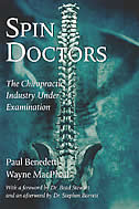
|
Spin Doctors The Chiropractic Industry Under Examination Paul Benedetti Canadians visit chiropractors about thirty million times a year, and surveys show that patients are generally satisfied with them. But Paul Benedetti and Wayne MacPhail have another opinion. Their hard-hitting CANOE.CA web site called Spin Doctors I & II were instrumental in educating the public about the excesses of some chiropractors. This book took years to write, and it is a must read for anyone who plans to go for chiropractic treatment, or who pays for insurance that covers it.
|
| 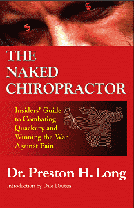 The Naked Chiropractor The Naked ChiropractorInsider's Guide to Combating Quackery and Winning the War Against Pain Dr. Preston H. Long Dr. Preston H. Long is THE expert. Consumers trust Andrew Weil for reliable information about alternative medicine, Dr. Bernie Siegel for inspiring words about mind-body connection, and Dr. Dean Ornish, for practical ways to keep their hearts healthy, but who the recognized authority on back care and the limits of chiropractic medicine? |
 |
|
Amani Oakley's closing in the Lana Dale Lewis InquestDecember 10, 2003THE CORONER: Ms Oakley, it's 12 o'clock. It's earlier than what we would normally take our lunch recess. I will, however, provide the option to you to either start now and we would take our lunch recess starting around 12:45 to one o'clock or we can take our lunch recess now and you may make your final submissions following lunch. MS OAKLEY: Thank you sir. I think I'd like to start now if that's okay. I can tell you as I turn to you now what kind of a relief it is to actually look you full in the face. All this time we've been sort of looking at each other from the side so it is, I think as all the lawyers have said, this is a moment where we get to actually look at each other for the first time through this entire inquest. Everyone else has thanked you. I need to say beyond thanking you, the family looks to you, the jurors as a source of its hope that many of the concerns that have been raised throughout to be dealt with. I also want to take a minute to thank Dr. McLellan because it has been an extraordinary inquest and his patience has been very appreciated. I have to say, I am going to apologize up front. I have a lot to say and I have exactly the same amount of time to say it as each of the other parties so I'm going to have to whip through things and I hope that with all of the knowledge that you have that it won't be too much of a problem. My position here is that if you go back to the beginning and I know this has been so very long so I think part of the problem here is going back to the very beginning, you'll see that the other side's strategy, the strategy of the chiropractors throughout this inquest has been to throw whatever they can at the wall and see what sticks. There's been a lot of theories that have been thrown out at that wall and have fallen by the wayside. We've heard about the heart. We've heard about haemoconcentration, overweight, high stress from alleged sexual assaults from childhood, her husband hitting her, hit by a car four months before, fell in the shower a year before. I know you remember all these things. We're not talking about most of them now but it indicates to you the procedure here was let's see what we can throw at this. Let's see what sticks. When all is said and done these theories put forward by the other side have crumbled. No basis in fact. No evidence to support them. Most of them nothing more than fantasies of lawyers in many instances of what can only be described as a real stretch. And of course this willingness of the chiropractic lawyers who switch to different theories, just so long as it wasn't related to the chiropractic neck manipulation injury was also seen in their experts and I'm going to show you that as we go through this. Even Mr. Schneider and you remember Mr. Schneider was here for a good long part of this inquest, made note of this fact on one occasion. Here's what he said December 12th, 2002. He said, "The only comment I make, Mr. Coroner, is that I heard Dr. Rosso change his opinion on the witness stand." I'm going to tell you also that the evidence that I'm going to give you and that the information I'm talking to you about, I have drawn so heavily on the actual transcripts because what you heard just now for example from Mr. Paliare, I will tell you a good percentage of that is completely in error. And I'm not going to tell you just what I think of the evidence. I'm going to tell you what people said. So that you know it's not my voice that's talking to you. It's the actual evidence. I've gone through these transcripts and I'm quoting. Mr. Foster, after we heard something like that from Mr. Schneider talking about the changing of the evidence of Dr. Rosso, and you remember Dr. Rosso. Dr. Rosso actually got in the stand and abandoned entirely the report he had made. The report he had made said it was the heart. He got on the stand, all of a sudden it was atherosclerosis. Not a word anywhere ladies and gentlemen for me to prepare, nothing. He walked into the stand, changed his entire testimony and then and then he says "Well I didn't know that I needed to write another report." I mean really, I don't think you need to be an experienced expert witness to kind of figure out that when you're getting into the witness box and you already have been asked to write a report ahead of time and you've now completely changed your mind on that report, that maybe a second report is in order. And it is kind of laughable to hear from Mr. Foster in his submissions on Monday that it was Dr. Deck who changed his theory. Oh my goodness, I have to say that's totally ironic. Dr. Deck wrote his report, in his report of December, 2001, Exhibit 17 in these proceedings. He examined all the slides. He determined, he still reaches the same conclusion as he and Dr. Pollanen originally reached, that Ms Lewis' death was caused trauma. Page seven of that report, it gives a list of 12 points setting out a number of findings that point him to the findings of trauma as the cause of death. On the stand this man innocently says, oh goodness, he says, "I've decided that of the 12 points I've given you, instead of number three being the most compelling to me to support the finding of trauma, I've decided that it's number one." Well did we get applause from the chiropractic lawyers? Did they stand up and say this is in the highest tradition of science? That Dr. Deck upon reflection decided that the list he'd already given us, he's going to point to number one instead of number three as his most compelling. No, that was seen as a terrible occurrence, Dr. Deck changing his testimony. Where's the testimony changed? I just know in this instance your commonsense is going to prevail. Just imagine for a minute an argument you're having with your spouse. You're saying to your spouse, "I'm really angry with you. I think you show a lack of consideration and why? Because number one you went out last night and you stayed out late. Number two, you didn't phone me and tell me you were staying out late, and number three it was our anniversary yesterday." And then as you're arguing you decide that it isn't the most compelling evidence of this lack of consideration that he didn't phone you. It's that it was your anniversary last night. You changed you argument because you've now decided that the thing that you're really, really angry about is that he didn't call you as opposed to it being your anniversary? No, of course not. On the other hand you get Dr. Pollanen, throwing his entire theory out the window and putting in a brand new one, and really with no self-respect whatsoever you have the other chiropractic experts wholesale abandoning their opinions as expressed in their reports and running after Dr. Pollanen's theory with such haste that they made serious, serious blunders that have never been made more clear that they had no idea. They didn't actually assess the evidence themselves. They were very busy supporting Dr. Pollanen. And I'm going to come back to that during the course of my submissions to you. So all these theories thrown against the wall with the hope that something's going to stick. Of course once everyone's finished jumping ship and leaving behind all the rest of the sinking theories, the only two competing theories that are left, is that she died as a result of trauma to her neck from a chiropractic neck manipulation or did she die from atherosclerotic disease? So okay, Mr. Oakley and I on behalf of the Lewis family, we knew the job that we had to do in this case. We knew the case we had to meet as we say in legal circles. We knew that in order to be able to succeed in convincing you, the jury, that our experts were correct that Lana Dale Lewis died as a result of stroke from trauma as opposed to any of those other theories, we needed to show you that the competing theories didn't make sense. For example, A, we needed to show you there was less than a 50 per cent chance that the heart was the source of clots. We needed to show you there was less than 50 per cent chance that the cause of the death was haemoconcentration. We needed to show you there was less than 50 per cent chance that her cause of death was from an alleged sexual incident that may have occurred more than 35 or 40 years before her death. We needed to show you there was less than a 50 per cent chance that the cause of death had anything to do with an alleged assault by her husband. We needed to show you that there was less than a 50 per cent chance that the cause of death was related to being slightly bumped by a car as she was crossing the street four months before her death. We needed to show you there was less than 50 per cent chance the cause of her death was related to a fall in the bathtub which occurred more than a year before. And finally, we needed to show you that Lana Dale Lewis did not suffer from stage six atherosclerosis. So we set out to ask questions and put forward witnesses who could address these issues one by one and we addressed all of these theories thrown against the wall by the chiropractors' heart theory. In the end who is proposing the heart theory? Nobody. That's one that fell down. Dr. Pollanen who was the first person to put forwards the possible heart theory, abandoned it entirely in his own testimony. He told Mr. Schneider that there was absolutely, sorry, I'm going to read this here. "It is highly unlikely that the clot originated in the heart and went into the posterior circulation." You also heard evidence, there was suggestions that well, if there was nothing wrong with her heart why wasn't the whole heart transplanted? Why just the valves and the aortic arch? But you also heard evidence there's a reason for that and we didn't get much into it because honest to God we have so much already to cover here. But in order to transplant a whole heart, the heart must be beating at the time that it is harvested, and that wasn't the case in Lana Lewis' case. In any event, as I say to you, which of the experts is still clinging to the heart theory? Nobody. Haemoconcentration was another ridiculous theory that held no water once examined. Frankly as well given the limitations placed the family's expert witnesses in supposedly testifying in areas that they were not qualified to testifying in it's amazing to recall the theory of haemoconcentration was put forward by Dr. Moulton and by the way, he's an orthopaedic surgeon. He is not, as you heard from Mr. Paliare, a neurologist. He has no expertise at haematology or conditions such as haemoconcentration since such conditions have nothing to do with what he did. Hopefully you recall what happened when Dr. Moulton was cross-examined by me on that theory. He admitted that (A), haemoconcentration was a condition whereby a person had too many cells in their bloodstream, those cells being white cells, red cells, and platelets. He also admitted, (B), Ms Lewis' cell counts were perfectly normal; (C), He admitted haemoglobin is not a cell but is a substance found within red cells and so an elevated haemoglobin level cannot form the basis for a theory that a person has haemoconcentration. He also admitted that, (D), he knew that people who smoked tended to have elevated haemoglobin levels. Every other witness who has been examined on this theory has confirmed Ms Lewis did not have haemoconcentration. So there goes that theory. As for the theory put forward by Mr. Danson that high stress levels from an alleged childhood sexual assault, I have to say this theory was nothing short of obscene. You'll notice that you heard no evidence, no evidence for this from any expert witness at all. The only evidence you heard about this was from Mr. Danson and you've heard the Coroner tell you repeatedly that us lawyers, we can't give you that evidence. There is no question that a lawyer has a duty to his or her client to represent them the best way they can. But they also have a duty to the judicial system to not turn proceedings into a circus. By throwing in entirely unfounded theories that an alleged childhood assault had anything whatsoever to do with Lana Lewis' death at age 45, Mr. Danson deliberately and cruelly smeared the reputation of the Lewis family, not to mention violating the requirement of the legal system to not identifying victims of sexual assault unless they want to be identified. In any case, I am certain that you will find that there was no basis laid for such an absurd theory. No experts adopted the theory and as a result the pain inflicted on the family was inflicted without any solid expert foundation and therefore for no good reason. Such a tactic by Mr. Danson could only have been directed at hurting as the family without any experts to back it, it had no chance of succeeding. Pretty much the same kinds of things can be said about suggestions made by Mr. Danson and Mr. Foster that Jim Sweeney hit Lana Dale Lewis and this is what resulted in her death. Great theory but once again smear the reputation of the family. Only one small problem again. No evidence. Not only that, but look at the ridiculousness of the theory in the big picture. We saw the letters Jim Sweeney wrote to the Coroner's office asking that his wife's death be investigated. Mr. Foster and Mr. Danson suggested that Mr. Sweeney had a guilty conscience in adding in one of his letters that he was prepared to take a lie detector test. At first glance, it certainly appeared unusual to say that, but his explanation is that he thought all options were going to be examined by a circle of experts by the Coroner's office and he wanted to let them know he would cooperate every way he thought possible. Mr. Danson, in his submissions, suggests that Jim Sweeney saying that he would take a lie detector is like a deer caught in the headlights, a confession of guilt. The only guilt Jim Sweeney had was being stupid enough to recommend that his wife attend a the chiropractor for treatment of migraine headaches. Thinking about it for a minute, the Coroner's office had done absolutely nothing in terms of investigating Lana's death as far as Jim Sweeney knew. At the time that he wrote those letters, pushing the Coroner's office to investigate, the Coroner's office had not been in contact with him at all, or given him any information to suggest to him that an investigation was underway. So what you're being asked to believe here is in a situation where Jim Sweeney had guilt about his involvement in his wife's death allegedly, he is the one that pushed the Coroner's office to investigate? He is the one that called and wrote letters saying what's going on? Please investigate? Does that make sense? What you're going to hear from me, is I'm going to tell you over and over again that whenever possible look for hard evidence that is not open to different interpretations by anyone. As far as I'm concerned if you're not convinced by what Mr. Sweeney said, that he didn't hit his wife, and that didn't cause her death, look at the hard evidence. It's easy. If Jim Sweeney hit her, the obvious thing to expect is there would be bruises on her body, both in her neck area and other places on her body if the two of them were involved in some kind of physical altercation. You heard evidence from various witnesses including Dr. Dhanani that if someone were to attend an emergency complaining about neck pain it would be an obvious that that an examining doctor would look to a neck to see if there was bruising and would definitely note it if there was. There's no bruising on her neck. It's not noted in the emerg records and it's not noted by the way in about five other places. September the 1st, which is the day that she went to the hospital, on page 529, there are diagrams and you'll see them if you look at the records in your deliberations. There are little stick figures, or little silhouettes of a body and they are found routinely throughout the hospital record. What they say is skin, on the one side here and it's got boxes for intact, abrasion, laceration, bruising, lash, ulceration, redness, swelling, raise, removed and so on. And under skin on September the 1st when she entered in hospital it says "intact." The box for bruising is not ticked off. That's what you call hard evidence. So we don't need to believe Jim Sweeney or not. Take a look at the hospital records and just in case there's a possibility that it was missed in emerg, take a look at the other pages where her condition for bruising is noted on September the 3rd. So that's second day in or third day she's in hospital. Page 539 of the record it shows there is a bruise, right loin from the angiogram. So there it is, it's actually X'd onto the little diagram I showed you on the right loin area, it says bruising. September the 4th, again she's examined for bruising. Page 549 of the records, again the right groin bruise is shown. Then September 10th, when she comes back to hospital, page 540, I apologize, 450 again the bruise on the right loin is noted. Nothing else. And September the 10th again on another page 483 it says they checked for bruising, none found. Then of course there is the actual autopsy record where the body was examined. The body was examined so thoroughly that you will see and the exhibit I believe is 16 that in the notes she has a bandage in the middle finger of one of her hands. So ladies and gentlemen if there was bruising you better believe it would have been picked up either in hospital in the many days she'd been examined or on autopsy, wouldn't it? Don't forget she went to see Dr. Knapp in the interim between the hospital visits. She told Dr. Knapp in the past about altercations she'd had with her husband. Why wouldn't she now? Why if that were the cause of the problem wouldn't she have said to Dr. Knapp, "Jim did it again. He hit me." There you go. And Dr. Knapp in fact indicated he would have expected that. She was not the kind of woman who wouldn't have told him. So how does any fair-minded lawyer continue putting this theory forward knowing the pain it's going to cause the family? Common decency ought to prevail when there's no evidence of any kind of supported theory that will be devastating to a man who still blames himself entirely for being the one who told his wife to see the chiropractor. Just another theory that the chiropractors threw against the wall hoping something would stick. With this theory like the previous one though, I think there was a hope that not only might some of the theory stick to the wall and maybe muddy up the waters, but they were also hoping that negative feelings might be generated against the family. Chiropractic lawyers want to whisper to you, this man beats his wife. This woman's father sexually abused her as a child. Shocking. Whatever the chiropractor did couldn't have been that bad. And I want to remind you that Mr. Danson never did abandon this theory. As late as Dr. Whitwell, Mr. Danson suggested to her that there was abuse from the husband and so in terms of what we were talking about with Dr. Whitwell the only thing that Dr. Whitwell would have had any interest in is if that abuse was known to have occurred around the time of death. The other evidence, things like being bumped by a car and falling in a bathtub, well I think they were so unsupportable that they were abandoned without much of a fight. So after leaving all these other theories pooling on the ground at the base of the wall with nothing sticking to that wall, we now come down to the theory of atherosclerosis having caused Ms Lewis' death. This is harder to dislodge off the wall but it is dislodged once you carefully look at the evidence that we've heard at this inquest. As I take you through the evidence I'm going to ask you to keep in mind the timing of events at this inquest. I'll point out to you what I mean as we go through the evidence but for now I need you to keep in mind different theories emerge at different times. We heard from a number of neuropathologists, pathologists and pathology witnesses who felt there was evidence of trauma in the slides they examined. The witnesses who thought they saw trauma in the slides they examined are, Dr. Pollanen originally, Dr. Deck, Dr. Whitwell, Dr. Fornasier, Dr. Richardson. The pathology witnesses who felt the slides did not show trauma, Dr. Pollanen, finally, Dr. Rhodes and Dr. Ramsay. The pathologists and neuropathologists who felt that the cause of death was trauma, pointed to a number of things to support their position and I'm going to come back to the issue of trauma but I first want to deal with the atherosclerotic theory. If you remember the first witness to put the atherosclerotic theory forward it was Dr. Michael Pollanen. Remember the tons of information you were given to justify this theory? As I recollect we've heard from all manner of witnesses who supported the theory that atherosclerosis killed Lana Dale Lewis and all of them took great pains to point that to you. Not the presence of atherosclerosis ladies and gentlemen. In fact we were told that the presence of atherosclerosis was pretty normal in everybody and atherosclerosis begins to make its appearance as early as age nine. It wasn't the presence that was emphasized to you over and over again. Instead in the case of Lana Dale Lewis the important thing to note about her atherosclerosis was it was severe and if you recall we were also told what severe meant. Let's go back to Dr. Pollanen for this. Dr. Pollanen spoke about six stages of atherosclerosis. The sixth stages as set out by the American Heart Association. Remember all that talk about stages. And he got the stages and he told us was from Robins. The pathological base of the disease that prevents the addition and he quotes the Bible of pathology and he says it was basic text that all pathology students are familiar with and have read and all pathologists are familiar with as well. And he described atherosclerosis in this way, and I'm quoting. "Atherosclerotic process involves and is very complicated but essentially involves the deposition of certain types of material and cells within the artery such that the artery becomes clogged off or occluded. There are certain risks for risk factors for that process but essentially it culminates in what the American Heart Association has classified as six different lesions. Stage one and I just draw your attention to this diagram" and this is Dr. Pollanen speaking, he is looking at the chart now, "and this shows stage one, two, three, four, five and six. Obviously six is the end stage. Type one lesion is the initial stage and this is generally correlated with other observations such as early onset in general terms as well as clinical correlation. So basically you have six stages. In the first decade of life the changes are minor consisting of small accumulations of fat, we call them fatty streaks, on the surface of the artery. And by the fourth decade, that's in the 40s essentially, if you're going to develop atherosclerotic disease what happens is" and listen carefully to this "the plaque becomes what we call complicated." That's Dr. Pollanen telling you what to look for in plaque. "But there wasn't a high degree of what we call stenosis" this is another area, another quote. Dr. Pollanen is talking about what he's seeing and what he's expecting. "The arteries are blocked by atherosclerosis. It's sub-occlusive. It's calcific, which means that calcium was being deposited into the wall of the artery as part of the disease process." Another thing to look for, calcium in the walls and he says, "If you remember back to the diagram I showed a table. The atherosclerotic process occurs over, over a period of time, years, and the end stage includes mineralization or deposition of calcium within the wall of the artery and that is a generative feature of this disease." Dr. Pollanen is answering questions from Mr. Schneider at this point. So Dr. Pollanen testified he looked for characteristics of stage six atherosclerosis and said that he found those characteristics in Lana Dale Lewis' plaque. Here's what he said he found. He said he found calcification. He said he found pre-aneurismal changes. He said he found ectasia. He said he found impingement on the lumen and he said he found neo-angiogensis and haemorrhage of the plaque. That's why he then said to Mr. Schneider, "I saw those things and I'm saying because I found those things she had stage six atherosclerosis." So even Dr. Pollanen and you're going to hear this echoed over and over, said to you it's not the presence of atherosclerosis, it's the characteristics and he lists what he says were the characteristics he said he found. I'd like to ask you because you now know so much about this process. Early on when we were hearing from Dr. Pollanen and Dr. Deck, we took their word for whatever they saw under that microscope. You said you saw calcification? Guess it was there. We just took their word. But we know now so much more and I'm now going to ask you to critically evaluate whether or not those findings actually were there. So there's a common theme that we've heard from every single person who told us about pathology, neuropathology and several of the neurologists as well, and that is, except for Dr. Ramsay, they all said to you in order to determine if atherosclerosis killed Lana Dale Lewis we must determine how severe her atherosclerosis was. As you could see, through this inquest, it was highly unusual to have a meeting of the minds on just about anything but they met on that. Except for Dr. Ramsay. Now, if you think back, what you will realize is that different experts use different language to describe this common theme. Dr. Pollanen talked about stage six atherosclerosis. Dr. Richardson determined that whether the plaque was safe or a vulnerable plaque and whether the plaque stenosed or closed down the lumen of the artery or the opening in the artery. In a few minutes I'm going to show you, despite using this different terminology ßeveryone's really saying the same thing. They were all saying: were the characteristics of atherosclerosis seen in Lana Dale Lewis' arteries consistent with a woman in her mid-40s dieing from atherosclerosis. There were slight variations on the same theme. For example, Dr. Pollanen looked at it this way. Were the characteristics of atherosclerosis consistent with stage six atherosclerosis and therefore responsible for the death? Dr. Richardson would put it this way: Were the characteristics of the atherosclerosis consistent with vulnerable plaque and therefore responsible for her death? The only expert we've heard from who said the characteristics of atherosclerotic plaque were not important was Dr. Ramsay. Dr. Ramsay told us the specific characteristics of the plaque were not in issue for him at all. He told us it was sufficient for him to simply see the plaque next to the thrombus and he would then equate the presence of thrombus to that plaque and call it a day. Ladies and gentlemen I'm going to ask you that you completely reject the position of Dr. Ramsay. Here's one of those issues of timing that I flagged for you early on. Dr. Ramsay appeared after all the pathology experts of the family were finished and after I had cross-examined Dr. Rhodes and showed that supposedly key findings made by Dr. Pollanen to support that atherosclerosis was stage six and severe were not actually present in the slides. Dr. Ramsay was even allowed to testify after Dr. Whitwell who rejected all of Dr. Pollanen's supposed findings that were meant to support a conclusion that atherosclerosis was severe. So after all that, we suddenly hear from a new expert of Mr. Danson. An expert who very conveniently now says, don't worry that all the supposed indications of severe atherosclerosis have now been shown to be non-existent. The final witness is brought in to save Mr. Danson's atherosclerosis theory by now saying that the characteristics of plaque don't matter a wit. He very simplistically says if atherosclerosis is there and next to a thrombus that's good enough for me. Let's everyone go home now. You know, I don't know about you but I find it really hard to believe that Dr. Ramsay was the only one of the all the neuropathologists and pathologists that we heard from in this inquest who knew what was going on. Dr. Ramsay actually said something that helped us here. He said look for areas of agreement. And ladies and gentlemen one of the key areas of agreement on experts on both sides of this debate is that they all testified that they needed to assess the characteristics of atherosclerotic plaques as seen in Ms Lewis' arteries in order to determine the severity of the atherosclerosis. We all know that the experts do not agree on their conclusions but there is equally no question that all the experts, except for Dr. Ramsay, testified that they needed to look at the characteristics of atherosclerosis, not merely the fact that it was present. So Dr. Ramsay suggested, look for those areas where the experts come together and I suggest to you the following experts came together to say the nature and characteristics of the atherosclerosis need to be determined: Dr. Deck, Dr. Pollanen, Dr. Richardson, Dr. Fornasier, Dr. Cheung, Dr. Rhodes, Dr. Whitwell, and Dr. Rathbone, all of them said the same thing. No one, except for Dr. Ramsay is sitting in the camp that suggests that those characteristics are unimportant and only the proximity to the thrombus is important. On that basis I think it makes sense to reject Dr. Ramsay as the odd person out on that. Once you reject his position that leaves us with a need to look over the evidence and determine what we know about the severity. If the experts only needed to find evidence of atherosclerosis and then they could all pack it in, well there really wouldn't have been any controversy because they all saw it so it's not that they didn't see it, they saw it. I want to caution you on what they're asking you to accept here. If you were to accept the proposition that just looking at a body and finding evidence of disease process then you can just go home, what you would basically be saying is that someone who has evidence of some other disease process in their body would never be found to have died from some other cause other than the disease process that they already have. That's clearly absurd. No body, except maybe the bodies of young children, is free of all disease processes and this is what has been so disturbing about this inquest. The chiropractic community seems to be saying to you that unless you have a perfectly healthy individual who is the perfect weight, who eats right and doesn't smoke and who is young enough they don't have appreciable atherosclerosis in their body, you can't conclude that a chiropractic neck manipulation was responsible for the death. And I say to you that the knowledge of what disease process may be found in a body and the assessment of whether that disease process killed the person or some external force killed the person is what pathologists and neuropathologists do all the time. For example, you may have heard a story on the news just last week about a very large black man who was beaten by police in Chicago and he died. Most of the police beating was caught on video. The man who was beaten weighed 350 pounds. On top of that he had cocaine and ecstasy in his system. The coroner who investigated the death announced in the news that he recognized both the fact that the man was dangerously overweight and had drugs in his system but the coroner still said that the cause of the man's death at this particular time was the police beating. Now what the chiropractors are asking you to do in this case is the equivalent of having the police take the position in that case, that since the man was obese and had drugs in his system well it couldn't have been the police beating that did it because he was dangerously overweight and he had all these drugs, that could have killed him anyway. Commonsense should tell you that you can still beat to death someone who is obese and you can still beat to death someone who has drugs in their system. A person may be in very poor shape, say someone who's homeless and doesn't have enough to eat and sleep out in the cold and still be beaten to death and a good pathologist should be able to see the effect of obesity, the effect of the drugs and the trauma and be able to figure out the cause of death and to screen out the background noise which is in all our bodies all the time and it's going on as we live and breath. In the case of Lana Dale Lewis what I need you to do is sift through the evidence to determine if the presence of atherosclerosis answers the question of what killed her or if it's simply background noise like this other man's obesity and drugs in his system. And Ms Vance, I remember a question of yours to Dr. Ramsay. In particular you asked whether he thought that Ms Lewis' arteries were so diseased that it had to be natural causes and of course he answered yes. But Ms Vance, I want to take all five of you jurors through what the experts told us about how to assess the severity of atherosclerosis in order to determine if Ms Lewis' arteries really were so diseased. One of the things that we were told is that severe atherosclerosis is characterized following a lot of calcium. Specifically here's what we heard on the topic. Dr. Pollanen, again he referred to Robins and as I indicated previously he says that the plaque becomes complicated and then he says it's calcific, which means calcium is being deposited into the wall of the arteries and he also talks about the atherosclerotic process appearing over a period of time and years and the end stage includes mineralization where the deposition of calcium was in the wall of the artery. And it's a degenerative feature of late disease process. To Ms Rothsteen, who was here for the College, he answered that he felt what his what he was observing, she asked was it calcific. He says, "Correct." Question, she says to him, QUESTION: And calcific is one of the indicators you told us of the severity of atherosclerosis, is that fair? ANSWER: That's fair. Dr. Rhodes, now Dr. Rhodes and Mr. Danson answering questions. Here is the question, QUESTION: All right and I just want you for the sake of clarity that you agree then with Dr. Deck and Dr. Pollanen's description of what you have just described? ANSWER: I think I used the same words. As I pointed out this to the jury I didn't mention the area of calcification. You see a little bit here, but remember the tissue has to be decalcified in order to prepare the slides. Calcium in sections like this stay blue and so part of the things, one of the things that happens when the atherosclerotic plaque degenerates is calcium is deposited." And to me, my question: "QUESTION: Presence of calcium is there, what's the explanation of the presence of calcium along that vertebral artery," I asked Dr. Rhodes. He says, "ANSWER: Definition of calcium in any tissue is a common degenerative change. It's not specific to atherosclerosis. Many, many diseases when tissue becomes damaged, one of the things that happens is calcium precipitates in the tissue. QUESTION: When you say it's not particular to atherosclerosis, is it something you would have seen in advanced atherosclerosis? ANSWER: It's a common finding in advanced atherosclerosis. So common that in fact it's used almost in the way we describe it. We would say advanced calcific atherosclerosis and grossly you recognize it because when you handle the artery it's crunchy. I mean you can't put a knife through it sometimes there's so much calcium in it and this is this is the normal course. This is what you see in advanced atherosclerosis." Dr. Cheung, I asked him about calcium and he told us in his presentation and answers to questions from me that he didn't see any calcium on the CT scan and he would have expected to see calcium on the CT scan because what a CT scan does is it will show bone and calcium is the thing in bone that makes things show up white on things like x-rays and CT scans. So Dr. Cheung specifically looked for that calcium because I said to him, well the theory on the other side is very severe stage six atherosclerosis, heavy calcification. And he said well if you slice the CT scan slices basically through the brain as it takes its pictures you basically see a bunch of rings if it was that calcified and it he said "I didn't see it" and he testified to that here. Dr. Whitwell I asked her and she answers, "ANSWER: When calcification is extensive it's best identified macroscopically. There's almost a crunchy crunchiness to the blood vessel. QUESTION: Would you agree with me that if you found blood vessels that were crunchy upon close examination that would be an abnormal finding and would be identified in the gross" and she said yes. QUESTION: So if it's not in the gross, one of two things, either an error has occurred or it's not there because there's no crunchiness that was found in the artery. ANSWER: Correct," she says. Dr. Richardson answered a question of mine and I put to him that quote from Dr. Rhodes where he says calcification is so characteristic of and Dr. Richardson agreed, he said "Yes, of course. In fact sometimes you could even see bone formation in vessel walls." "QUESTION: So are you in agreement that if this is severe atherosclerosis you would expect to see heavy calcification? ANSWER: Yes. QUESTION: And you don't see any with Ms Lewis? ANSWER: I don't see any. QUESTION: Okay, and Dr. Rhodes talked about the fact that the artery was crunchy in severe atherosclerosis on gross. Is that something that you agree with? ANSWER: Yes." So groundwork, one of the characteristics we've been told by all these experts to look for is presence of calcification. Now question, was calcification present in Ms Lewis' artery? So in terms of a list of questions you need to ask yourself when you sift through whether or not the evidence supports the severe atherosclerosis, that's one. And I am certain and it's rightly so, your attitude is going to be, well she's going to tell us what her expert said and not surprisingly her experts are going to agree with her. So what you're going to see that I do repeatedly for you ladies and gentlemen is I'm going to show you what the other side said because I want you to understand that this is not just from my side and not just from my experts. So, and the other thing I'm going to do is I'm going to reference back to hard evidence again, and one of the things that's hard evidence in calcification again is it wasn't seen on CT scan. That's something that you don't need to rely on someone else's interpretation. It's white and Dr. Cheung said it would show up white and he showed you the CT scan and he said it's not there. So with your own eyes you could see that it's not there. There wasn't any white. And Dr. Rhodes what did he say about calcification? If you'll recall I caught Dr. Rhodes saying some things that aren't at all correct. You might recall this exchange between Dr. Rhodes and I. "QUESTION: Well actually, what I wanted to ask you was about your testimony with respect to calcium. You've indicated that despite the fact that you didn't see a lot of calcium, you're concluding that it must have been there because you saw some bits of blue remaining and your testimony was, if I'm not mistaken, that a sample of the tissue was decalcified. ANSWER: I have a great deal of experience with decalcified tissue. I work with it all the time so I know from my experience where I handled the gross tissue and then looked at the slides that once you decalcified it, most of the calcium disappears. That's what you're doing when you decalcify. So I have a great deal of experience saying that after decalcification I see just a little bit of calcium left and extrapolating from that to what must have been there, before you decalcified it." And so I ask him, "QUESTION: So you're extrapolating from what you're seeing? I want to be clear on this. And he says, "ANSWER: You asked me whether there was calcification, or someone asked me, and my answer to that was, the answer is yes. You asked me how I knew and I said because even the decalcified artery, there was still some calcium remaining. And then he says, "ANSWER: I'm just telling you that once you decalcified it, it won't be as blue as it would be if you didn't decalcify it. So I've got a bit of blue, blue" is what he says, "I know there would have been much more blue had we - they not decalcified the tissue." "So you're extrapolating" this is what I say to him, "and you say there would have been much more blue if it hadn't decalcified is that right?" And he says, "That's correct." "QUESTION: Okay, so that's your position is that you're extrapolating from what you've seen and your submission on that was it would have been much more blue. And he says again, "ANSWER: You see right now a little bit of calcium in the decalcified tissue. You know there would have been more before you decalcified it." He also answered one of Mr. Danson's questions and he says the same thing. He says, "You see a little bit here but remember the tissue had to be decalcified in order to make slides. Calcium in sections like this stays blue. And so one of the things that happens with atherosclerotic plaque as it degenerates, the calcium is deposited in to it." Dr. Rhodes seemed to forget what other experts clearly told you: no one decalcifies the brain. We heard that from Dr. Richardson and we heard it from Dr. Whitwell and you'll notice when Dr. Ramsay came to testify, he didn't tell you that we were wrong on that. Use your commonsense. They all told you what decalcification was for. It's to remove calcium from bone. They also explained what happens in an autopsy. The brain gets taken out of the skull. There's no bone. They also told you the brain is very soft tissue. Putting it in a tough solution, acidic solution like what happens in decalcification would mush the brain entirely. It's never done. And so I ask this of Dr. Whitwell. "QUESTION: Dr. Whitwell I just want to confirm with you in terms of the brain, the brain was separated out from the body and it is not decalcified, is that correct? ANSWER: Correct. QUESTION: You never decalcify the brain. Is that correct? ANSWER: Yes, that is correct." QUESTION: So whatever calcium you see on the slides is all there is. It's not a matter that some of the calcium has been removed from the process of decalcification. Is that correct? ANSWER: That is correct. And then nobody raised it, so now you know what Dr. Rhodes told you he saw. He saw a little bit. He described in fact to Ms Jones as a hint of blue. That's the amount of calcium that was in Ms Lewis' artery. A hint of blue. And contrast that with what they all told you they need to see if it's severe atherosclerosis, serious, severe, lots of calcification. Dr. Rhodes saying to you if in fact you call it you call it in the name, you mention calcification because it's so much a characteristic of this disease. So here's where he says to Ms Jones, "ANSWER: That means the atherosclerosis itself didn't block off the artery. It's calcific. You remember it was hard to see because the calcium's been removed, but there was, there was a hint of calcium. QUESTION: And that's the blue you pointed out? ANSWER: That was the blue that I pointed out, exactly right. ANSWER: Most of the calcium is removed, as you decalcify the block you stop after you get to the point where you can cut the knife through it and that leaves a little bit of calcium. As I showed you in the slide there was a faint hint of blue on the top there so I can't see most of the calcium because most of it's gone but there was there was a faint hint left where there had been some." Now I don't want to confuse you. The decalcified portion, and you'll see this in an original autopsy report, is the neck. So what happens is after they go in and they get that brain and they snip the arteries, the vertebral arteries and the carotid coming into the brain so they can lift the brain out. What's left, the arteries as they work their way through the bone in the neck, that's the part that's got to go in solution because they can't slice through that with the bone in there mixing up with the arteries. But the brain part, no. And of course nobody saw atherosclerosis extra cranially so what we're talking about was all inter-cranial and that was never decalcified. And Dr. Whitwell, I asked her a question about whether she saw severe atherosclerosis and she again mentioned that the best place to see severe atherosclerosis would have been in the gross and she says yes, it's crunchy. Yes, I would expect that it gets reported if it's crunchy and she confirms what you heard from Dr. Richardson and Dr. Ramsay that you can see these nodules in the tissue, there's chunks of them and she confirms that's an abnormal finding and yes she'd expect to see it on gross and no you don't see it on the gross here. And then I ask her specifically, you know I went through the whole list of Dr. Pollanen's observations that he says he makes in support of a finding that she has severe atherosclerosis. I went one by one by one with those points with her and the second point was that Dr. Pollanen says he sees micro-calcification. Actually he's very clever because of course what Dr. Whitwell said to me was, well, micro-calcification implies there are very small amounts. Remember the big words Mr. Paliare said to you today? These experts used too many big words, too many scientific words. Dr. Pollanen was very good at using those very big words. So instead of telling me there was very little calcium he called it micro-calcification. Same thing. Virtually nothing there. And Dr. Richardson, I asked him what he saw and he said he saw two little specks of blue that I think were calcium that that's all. Otherwise he said no calcification. So what I think you can take from this evidence ladies and gentlemen is that: one, serious calcification is a characteristic of stage six or end stage, or vulnerable plague, whatever you want to call it. What ever we have heard from different experts. Atherosclerosis, in other words atherosclerosis that might kill you. You find serious calcification in that. Dr. Rhodes in fact told us one of the things that happened is that when plaque degenerates and calcium is deposited and he also told us that it's such a finding of severe calcification is so common that it is part of the name of the disease. Number two point you can take from this, Lana Lewis arteries contained a faint hint of calcium. Number three, that is not consistent with stage six or end stage atherosclerosis and is not consistent with level of the disease that could kill a person. Now, I see that it is five to one and what I plan to do after lunch, just so you know is I'm going to take a look at the other characteristics that we've heard also form a basis of a finding that atherosclerosis is severe. So that's the first one that I've done with you which is calcification and it is five to one so I think it is an appropriate time to stop. THE CORONER: Very well, Constable Drummond we will recess an hour and 15 minutes.
R E C E S S
U P O N P R O C E E D I N G: THE CORONER: Ms Oakley? MS OAKLEY: We ended before lunch on one of the points of Dr. Pollanen and all the pathologists in fact told you to look for if in fact a person has severe atherosclerosis and that's calcification. I took the opportunity to put Dr. Pollanen's exhibit up, Exhibit 36, because I think again it's very important again for you as you are going through the testimony that was heard here whether or not in the end you believe that the evidence supports the findings that Dr. Pollanen has drawn into that diagram and which many, many of the experts came in and said "I agree with that diagram." So just so that you understand: If those findings that underlay Dr. Pollanen's diagram are incorrect, and if the other experts then come in and say "I agree with that," they're wrong too. The next point I want to bring up in terms of what you find in serious atherosclerosis, and we heard about this, is ectasia. Dr. Pollanen testified he saw ectasia and he drew it into his diagram, that's the wavy line on the left side. He indicated that a finding of ectasia means atherosclerosis was advanced. Here's what he said to Mr. Schneider on this issue on May the 7th. "QUESTION: Now, what I wanted to ask you was, I assumed that if we're seeing ectasia then the this atherosclerosis is fairly advanced. Am I correct? ANSWER: Yes." And once again on the issue of whether ectasia is present or not, Dr. Rhodes is particularly helpful although probably not in a way that he wanted to be. As you may recall I asked him about his observation about ectasia. I asked, "QUESTION: And you said that you'd be looking for a similar calibre. You remember" this is me speaking, not Dr. Rhodes "that ectasia is of the calibre of the artery has now turned into sort of like a wavy line, there's areas out and in. It's the calibre, it's not even" so I asked him, "You said you'd be looking for a similar calibre. Can you show me where in your records where you've measured the calibre along the length of intracranial vertebral artery? ANSWER: Now this is best judged as you're looking at it and would have been best observed by Doctors Pollanen and Deck as you just look at the artery and you can look at it grossly, not under a microscope when you've got this piece of tissue, this tube." So he says if you see ectasia you'll see it on gross. He then admits that there's no observation of ectasia on the gross and you guys can see that with your own eyes. There's that hard evidence for you, back in Exhibit 16. I then take Dr. Rhodes' own words where he says ectasia is best observed grossly and this is want happened next, "QUESTION: Well, what I'm asking you is what you just said about the gross examination of the vertebral artery and that Dr. Deck and Dr. Pollanen would be in the best position in order to see whether or not there might be a ballooning out in some portions of the vertebral artery, and now correct me if I'm wrong, but the only place you could assess the gross observations made by Doctors Deck and Pollanen would have been in the autopsy report" and he says "Well I will correct, because you are wrong. The easiest and best way to look for some sort of dilation like that is when you've got the gross specimen but you can certainly recognize the ectasia looking at a microscope slide because once again you'll have an opportunity to judge the size of the vessel as in comparison to the thickness of the walls and so you can look for damage to the wall where it's abnormally thin and dilated. So you certainly can recognize it at a microscopic level. It's easier if you start with a gross examine." So basically Dr. Rhodes ends up saying the best place to look for ectasia is grossly but you can also see on the microscope slides. Okay, so we know that there's no mention in the gross description made by Doctor Deck and Pollanen and yet I do want to remind you that early on Dr. Pollanen told us that in his differential, in other words the list of things that he was looking for when he examined Ms Lewis' specimen both grossly and under a microscope, his list included atherosclerosis. So it's not as if that wasn't in the head of Dr. Pollanen and Dr. Deck from the start. So they're thinking atherosclerosis, they've got the gross, they'd be looking for these things. And remember one thing here about the microscopic examination. You heard from many of the pathology witnesses that the vertebral artery was not embedded properly in the blocks. The artery was embedded at an angle and this would certainly interfere with looking at the slides microscopically in order to determine the width of the artery lumen and the artery wall. Here's what Dr. Richardson said about this, "Well to evaluate the lumen size of the tube such as this or a vessel you must have sections that are absolutely true at right angles to the wall. If you cut them on an oblique angle and you just look at one section and everything is distorted, this is what's referred to as looking at it en fas (ph), en fas meaning you're looking at it all spread out and it exaggerates the thickness of certain layers. So to do it, you have to have absolutely pure cuts that are at right angles to the access of the vessel. If you don't have that, then it becomes very difficult to evaluate the lesions in the wall." So I'm going to charitably suggest to you that the reason Dr. Pollanen who was a trainee at the time that he examined these slides and he did not have as much experience as Dr. Deck and Dr. Whitwell and Dr. Richardson and Dr. Ramsay, the reason he sees what he believes to be ectasia is because he does not have enough experience to compensate for the poor placement of the arteries and the blocks. You'll notice that I included Dr. Ramsay in that group. He doesn't see any ectasia, either, ladies and gentlemen. You go back to Dr. Ramsay's exhibit which is 255. He went slide by slide. That was his big claim to fame. He gave us an individual description of 507 slides he looked at. You look at that list. No mention of ectasia. What did the other experts say about ectasia? Dr. Richardson said to me, I asked a question, "QUESTION: Now in diagram Exhibit 36, Dr. Pollanen has also drawn the left vertebral arteries having a wavy in and out appearance and he described that as ectasia and I'm wondering, Dr. Richardson, whether you saw ectasia in your examination of the slides? ANSWER: No I didn't. I believe last time when I demonstrated this was in a serial of block or series of sections of the artery shows there's no ectasia and the calibre of the reference was uniform throughout." And then I asked him, "QUESTION: Again we heard from Dr. Rhodes that ectasia can also be seen grossly. Is that correct? ANSWER: That's correct, yes. QUESTION: And is ectasia an abnormal finding? ANSWER: Yes, it means that somewhere along the line the vessel wall has been diseased or something happened to it. So instead of being the same calibre it's now dilated. If this is just uniform it's called ectasia. If it's unilateral, if it's just off to one side for instance, it's called aneurismal, an aneurysm formation and these are very dangerous to have in blood vessels in the brain so of course you'll note it. QUESTION: So on gross if you saw a vessel that appeared to have these ballooning about you would make a notation in gross that ectasia was present? ANSWER: Yes, that's part of the gross examination of the brain since the vessels are so vital to the brain. QUESTION: Now Dr. Rhodes actually indicated in answer to one of my questions, he said 'The question of whether or not there's ectasia present and where it's present is such a minor point that I did not look for it,'" and I asked Dr. Richardson, "Would you agree with him that the presence of ectasia and where it's situated is a minor point? Do you agree with him? ANSWER: No. Anywhere you see in a person of this age in their 40s and 50s and even 60s indicate it would be abnormal." Then I asked Dr. Whitwell the same kinds of questions, "QUESTION: Did you find evidence of ectasia as you went through? ANSWER: No. QUESTION: No, and is ectasia something else that can be seen on gross examination as well? ANSWER: Yes. QUESTION: Would it be considered an abnormal finding to be reported? ANSWER: It would, yes. QUESTION: And you don't see it microscopically either? ANSWER: No." And neither Dr. Deck nor Dr. Ramsay make any mention of seeing ectasia either with a microscopic examination of the slide. So with regards to the second point, this is calcification we've done, now ectasia. Regards to ectasia you can take from the second: number one, ectasia is a characteristic of stage six or end stage atherosclerosis. In other words it is a characteristic of the kind of atherosclerosis that might kill someone. Dr. Pollanen said that the presence of ectasia means atherosclerosis is advanced. Two, the best and easiest place to make an observation that ectasia is present is in the gross or visual examination of the vertebral artery. Three, no such observation was made about the presence of ectasia and Lana Lewis' vertebral arteries. You would expect that such an observation would be recorded in the gross because ectasia in someone in their 40s, 50s, and even 60s would be an abnormal finding and Dr. Rhodes told us that it is obvious that one would expect that a pathologist would record a normal finding. Four, the majority of experienced pathologists in this case and the ones who you can be sure are aware that the arteries were not properly embedded did not find any evidence of ectasia in Lana Lewis' vertebral arteries on microscopic examination. Five, that is not consistent with stage six or advanced atherosclerosis. Next what we heard from Dr. Pollanen that another characteristic that severe and advanced atherosclerosis is a lipid core and inflammatory cells. We heard about the presence of a lipid core and inflammatory cells in vulnerable plaque or advanced atherosclerotic lesion first, not first, but from Dr. Richardson as well. He brought with him a series of articles from Nature Medicine which were published in November of 2002. These articles are Exhibit 246 and one of the articles that Dr. Richardson referenced in this paper is entitled Vascular Proliferation and Atherosclerosis. The article starts out by saying, "Here we will review the current understanding of the patho-physiological mechanisms and the status of molecular and gene therapeutic approaches in vascular proliferative diseases. The understanding of the patho-physiology of atherosclerosis and related vascular diseases has changed over the last decade providing new perspectives for preventative and therapeutic strategy." On page 1250 of the article there are a series of four diagrams showing what one would expect to see at each stage of atherosclerosis. The first diagram shows the initiation of atherosclerosis; the second is entitled "an early lesion" and it also shows the presence of foam cells; and you may recall what Dr. Pollanen said about that. He indicated that first in atherosclerosis you see what's called a fatty streak and that's consistent with what you see in this Nature Medicine article. In the third diagram, labelled "vulnerable plaque" it shows the presence of a lipid core which is made up of abundant foam cells along with "intense inflammation" and the diagram shows that this inflammation is made up by the presence of lots of monocytes which are also known as macrophages and also T-lymphocytes. You may recall when I cross-examined Dr. Ramsay about these characteristics of safe and vulnerable plaque he first admitted that the last time he studied anything about atherosclerosis was back in school and he also said about Dr. Richardson's description of the stages of atherosclerosis, he says "nice theory." I'm sure you remember that. He suggested this was all new stuff, just theoretical and he even suggested that because the article had been written with the backing of drug companies, question mark, perhaps that was supposed to suggest that it was somewhat self-serving and suspicious. In keeping with what I'm trying to do for you by showing you that it's not just family witnesses that gave you specific information let's go back to what Dr. Pollanen told you about atherosclerosis. He referenced the book again by Robbin, which he called the bible for pathology students, and Dr. Richardson actually addressed Robbins and he said it's pretty much, it's a very elementary textbook and he said the whole science of atherosclerosis has changed an awful lot since Vercow (ph) and the current concepts are those that I believe are distributed in the review article from Nature Medicine. However, even keeping in mind that Dr. Richardson said Robbins is very basic, if you look at Exhibit 37 which are the pages from Robbins that Dr. Pollanen put in, he spoke about a chart and the chart was the one with the six stages of atherosclerosis. If you look at the chart, lo and behold, under type five lesion you'll find that it says that there's a lipid core or multiple lipid cores. As a matter of fact the chart shows the plaque develops from type one to type six lesions and it talks about the fact that there's a core of lipid that's developing and progressing and then it talks about, at stage type six a surface defect. So there's Robbins, this basic elementary pathology text Dr. Pollanen describes as a bible for pathology students and it's describing some of the same concepts that Dr. Richardson spoke about which you find in the Nature Medicine article and then you have Dr. Ramsay saying "nice theory." Well didn't he read Robbins? It's not new. According to Dr. Pollanen it's something every pathology student reads. The other thing that that the article and what Dr. Pollanen told us is that without the lipid core what you have is a simple plaque and he said that as a plaque develops and I think I read this to you earlier, it turns into what's called a complicated plaque and he claimed he saw complicated plaques in Lana Lewis' arteries and that was the basis for his finding that it was advanced atherosclerosis. So you've got the simple plaque and the simple plaque doesn't have things like the lipid core, and then you get the complicated one and it's got lipid core and that's what Robbins says. Now, I put this "nice theory" comment to Dr. Rathbone and I asked him if he'd had an opportunity to read the Nature Medicine article, and whether he was familiar with it and he says it's similar to what's been described in those articles but those reviews, those articles are reviews he says. "This is material that I knew about in 19 in the late 1960s, studies being done. We have perhaps the largest thrombo-embolism group in Canada, certainly maybe even the world, at McMaster led by Fraser Mustard and I was fully aware of the studies on blood vessels, platelets, and removal of surface material. So yes, this material I was aware had been published many years ago. More detail has now been added, we now found new proteins. We're filling in bits, but the actual information that we have discussed thus far as been known for about 30 years." No new theory. Then I ask him, "Are you familiar with concepts in the article of vulnerable plaque versus stable plaque?" And he says, "Yes. If the plaque forms and this is the fat deposition that changes and I won't go into the cells unless you want me to, that form these, the fatty cells that form this plaque on the wall of the arteries they get gradually covered over and as you can see in that picture of the article" and he's at this pointing to the Nature article, "there's a fibrous covering and then the endothelia cells line over this. Now if this covering is thin there is a distinct possibility that the plaque will rupture. If that happens platelets are exposed to these to the material inside the plaque, which the platelets see as a wound and platelets are set up to stop bleeding, so they form this plug on the vessel wall. So if the plaque is vulnerable it has a thin layer on it and it can rupture after a while and certainly we now know that since at least 15 years of studies have been done to show that if you lower the cholesterol by diet, by extreme diet or by various drugs such as Statins (ph) or if you lower the blood pressure one can get changes within these within these plaques so that the protective layers over the very reactive material is just thickened and so the plaque becomes quiescent and so they don't pose as much of a risk. That's why we don't recommend taking out all plaque out of the carotoid." In other words, you heard from a lot of the doctors that they don't go in and do surgery unless you get this really severe narrowing of the arteries and both Dr. Cheung and Dr. Rathbone said that even with a severe narrowing, unless it hits about 90 per cent, he also would be looking for symptoms. So the reason you don't go in I mean, you and I might think I certainly would have thought that something like 70, 80 per cent you might get a surgeon saying better get in there, but in fact what Dr. Ramsay says you don't go in there because most of the time that plaque is just sitting quietly and so you'd actually do more damage to go in. I'm sorry, I think I said something other then - I said Ramsay instead of Rathbone, which I expect to do a couple of times today so I apologize. There are a lot of Rs in this case. I don't know how it happened. And then I say to Dr. Rathbone, "Two days ago we heard from Dr. Ramsay, a neuropathologist and when I asked him about the vulnerable verses stable plaque his response was something like, 'it's a nice theory.' Is that what this is in your understanding? Is this just a theory that there is vulnerable and quiescent or vulnerable and stable plaque?" And he says, "No, it's supported by a huge amount of evidence over the last 30 years and this is something that's born out in clinical trials. It's born out in laboratory situation and it's the basis on which neurologists make their decisions about how they're going to treat patients with cerebral-vascular disease." So the Nature Medicine article indicates that a problematic or vulnerable plaque will have a lipid core and the presence of certain cells and cells are foam cells, monocytes, microphages. Now let's see what the experts said about the presence of these things in the plaque of Lana Lewis. I asked Dr. Whitwell, and her answer to me about what she saw in the plaque, she said, "In fairness most of the well I saw atheroma. I probably for oversimplification described it as atheroma but generally pretty simple plaque." Remember the simple plaque, stage one, stage two. Now you remember what Dr. Pollanen testified, he testified that atherosclerosis advances in stages. It goes from fatty streak, to simple plaque, intermediate and a few more steps and you get into the complicated plaque. Dr. Whitwell didn't see any complicated plaque. She saw generally pretty simple plaque. I asked her a question about what she had written into her report. "QUESTION: So all the plaque you described I do not see you describing any of the cells as monocytes or foam cells or fibroblasts inside the plaque. Would you agree with me? ANSWER: That's correct. QUESTION: Dr. Richardson we heard from indicated that the plaque was there but by and large was acellular. There were no cells in the plaque. Would you agree with that finding? ANSWER: I think that is a fair comment generally." Dr. Richardson told us that the plaque in arteries was pretty much entirely acellular and you remember Dr. McLellan at one point wanted me to be clear on what I was saying because the word is a single word, "acellular" with the a in the front, meaning no cells there. And he explained what it meant, that there are no cells in the plaque and he told us that cells have a nucleus and when they're stained, the nucleus stain is dark purple. So again, ladies and gentlemen I ask you, you have photographs that have been put as an exhibit. We showed them to you at the time but you're free to look in the jury room again and see if you can see the sign that Dr. Richardson said there are no nuclei, no dark purple spots inside the plaque. It is acellular, there are no cells in there. And again, I'm going to take you to Dr. Ramsay's report, not my witness. Exhibit 255. He mentions he sees focal, and we've heard that means in a spot, sparse mineralization, and that's the calcium, and collections of foamy cells in one slide. One in 507 that he described. And you can find that one slide in page nine of his report. Dr. Richardson and Dr. Whitwell both say that they do not see any lipid or fat collections in the plaques. Dr. Ramsay's report does not identify any slides that have any lipid or fat collection. So there's the lipid core that everyone's telling you to look for including Robbins, including Dr. Pollanen, including all of the other pathologists. They said they've got lipid cores. Well, you take a look ladies and gentlemen at any one of the pathology reports we have entered in as exhibits and you shall find nobody has reported a lipid core observation anywhere. And again, the inflammatory cells. You find this little pocket, a slide in A where they found some foamy cells, that's it. So with regards to what you find inside a vulnerable or advanced or complicated plaque, you can take from this evidence that: one, a lipid core and the presence of inflammatory cells are characteristic of stage six or end stage or complicated plaque or vulnerable. In other words again, the kind of plaque that will kill someone. Two, the evidence from Dr. Whitwell, Dr. Richardson, Dr. Ramsay is that there is no lipid core and only a rare presence of cells in the plaque in Lana Lewis' vertebral arteries. Three, the absence of a lipid core and inflammatory cells means that the plaque in Lana Lewis' vertebral artery was not complicated plaque. Dr. Whitwell called it simple plaque. Dr. Richardson called it safe plaque that was not in danger of rupturing and causing the creation of thrombus. Plaque which is not complicated and is not the kind of plaque that would rupture is not the kind of plaque that would result in someone's death. Another observation that Dr. Pollanen tells us to look for in serious or severe atherosclerosis is neo-angiogenesis and plaque haemorrhage. Dr. Pollanen says that these are also characteristics of advanced atherosclerotic disease. He answered some questions about them and here's what he said. "QUESTION: Okay, neo-angiogenesis means" and this is to Ms Rothstein "ANSWER: Proliferation of small arteries within the atheromatic element", in other words inside the plaque. "QUESTION: So again something you'd expect to see in end stage atheromatous? ANSWER: Yes." Now to Mr. Schneider on May 7th on pages 72 and 73 of the transcript, what he says to Mr. Schneider is I did see neo-angiogenesis but not at the site of haemorrhage. Now he says the explanation for the haemorrhage is the neo-angiogenesis. He then admits though that that's not where he saw the neo-angiogenesis and you won't see it there. Dr. Ramsay's report again comes in quite handy. Again he goes slide by slide, tells us what he sees. No mention of neo-angiogenesis anywhere in the report. Back to Robbins, they also indicate that a complicated plaque has surface defect of the plaque and haemorrhage of the actual plaque and both Dr. Richardson and Dr. Whitwell testified that there was no connection between the haemorrhage that they saw in the vertebral artery wall and any plaque. And by the way just because there are some red cells in the plaque doesn't mean that it's associated with the plaque. And I can just use a simple example. If you cut your finger and the blood runs down your hand you're going to have blood on your palm, but your palm isn't bleeding so it's the same thing. If there's blood in the artery walls it's going to bleed into the artery walls and just because some of the blood makes it into the plaque doesn't mean the plaque ruptured and in fact that's exactly what Dr. Whitwell says. "ANSWER: In fairness it is haemorrhage although whether or not it represents intra intra plaque haemorrhage which is generally a large amount of haemorrhage, what I did not see was a large amount of haemorrhage. QUESTION: So the haemorrhage can in fact come from tissue around the plaque and it could have some bleeding into the plaque? ANSWER: That is correct. QUESTION: And that doesn't mean the plaque itself is necessarily the source of blood, it's just the surrounding tissue is bleeding and going to bleed into in the vicinity? ANSWER: Correct." You'll see in Dr. Whitwell's report she makes the same conclusion. She says "Yeah, I saw some blood in the plaque. I don't believe it's connected to the plaque." So her observation is a couple cells of blood, not connected to the plaque. So again with regards to what you find in advanced atherosclerosis you can take from this evidence that one, neo-angiogenesis and plaque haemorrhage are characteristics of stage six or end stage atherosclerosis. In other words again, atherosclerosis that you might expect could kill someone. Two, the evidence from Dr. Whitwell, Dr. Richardson, and Dr. Ramsay is that these characteristics are not present in Lana Lewis' vertebral artery. Three, the absence of these characteristics means that the atherosclerosis in Lana Lewis' vertebral arteries is not severe, it's not stage six. Now another thing that Dr. Pollanen says is roughened surface of plaque, reduction of lumen sign. Dr. Pollanen explained that plaque might cause a reduction in lumen size in an artery and that the plaque can cause the surface to be roughened and this might cause clotting. We heard from Dr. Cheung, Dr. Rathbone, and Dr. Richardson and others that a closing down of the lumen of the artery or the opening in the middle by atherosclerosis is not of any real concern until the opening is down significantly, 70 per cent or more. Well actually Dr. I'm going to get into the numbers I guess. And I asked Dr. Rathbone when he would send a patient to a neurosurgeon for surgery with occluded vessels and this is what he said. He said, "I refer patients to a neurosurgeon who has atherosclerosis particularly of the carotid arteries. But for the stenosis, the narrowing of the arteries, the most important indication is that the person has some transient warning stroke,T-I-A in the territory in which there is narrowing. Now we look again at the narrowing and we take not just the extent of the narrowing unless it's very high and by very high that's" sorry I'm just going to pull what he was talking about at the time. He was looking at that photograph which is Exhibit 249 in these proceedings and you'll remember that this is a photograph from Greenfield and the top one shows lumen encroachment and he told us that the dark circle is the original artery and that the layers that you can see building up is the atherosclerosis growing into the lumen. So he said, he looked at this and he said, "The photograph that would be just bordering on very high. We would have to usually surgeons don't operate unless it's over 90 per cent stenosis if it's not symptomatic." "So symptomatic is very poor evidence and they do better with surgery but once you become symptomatic and surgery may help if the symptoms have been recent. But one thing we look for is evidence on the ultrasound and on angiography of an ulcer forming on the plaque. So the plaque is no longer smooth but there is some roughening of the surface." So according to Dr. Rathbone, a roughening of the surface of the plaque is the result of a formation of an ulcer. And Dr. Whitwell responded, when I asked her what would you expect to see in serious, severe atherosclerosis on gross. She said you would see ulcerated plaque grossly. So you could see the ulcers on gross. And again nothing like ulcerated plaques or lumens or openings of the plaque were seen on the examination of gross and Dr. Whitwell indicated that. Dr. Ramsay also doesn't note any observations about the serious reduction in the lumen of the vertebral artery nor any evidence of ulceration of the plaque. Dr. Willinsky, in describing what he saw on the right side of the angiogram, said it was smooth not roughened, which Dr. Rathbone then indicates would mean that on the right side where you're hearing evidence that she had serious severe atherosclerosis on the right side as well, on the angiogram what Dr. Rathbone says is, if you've got a roughened surface you see it on the angiogram, and Dr. Willinsky testified he saw it smoothly. So even if there's atherosclerosis on the right side, what Dr. Willinsky said is it's not ulcerated plaque because it's a smooth surface. Dr. Cheung also told us that even if you get clots clinging to the wall of the artery you would never get a complete occlusion like what we see on the left side here unless first you have serious stenosis with plaque. Think of this like a flowing river and you're not getting a plug in that river unless you first have an awful lot closing down that river. So in other words water's rushing along, you're going to get branches and and all kinds of debris sticking along side of that river but that's not enough to close a river and what Dr. Cheung said is even if the plaque was causing any kind of clotting along the side you also need this serious stenosis of the atherosclerosis closing down the lumen. So in combination, you get the serious stenosis, then on the part that's remaining you start getting clots there and then you can have it close right down and that just makes sense. You've got it wide open. It's not something they're going to occlude, like what we see in her left side, and that's what Dr. Cheung told us. Impossible, he said. And again, here's the hard evidence I'm going to ask you people to look at. You I invite you look at every photograph that has been entered in as an exhibit. I challenge you to find any impingement of a lumen that even comes close to this. You will find some impingement which is very minor. It just comes out, like, if you have to put a number to it, ten per cent, 15. You're not getting anything that looks like this in Lana Lewis' arteries. So there's your hard evidence again. Don't take my word for it. Don't take what the experts are saying. Go look at the pictures. This is Exhibit 248, Block A, and it's just an example of the many photographs that are in these materials. And I just want to be clear. This is all clot. You know that because you've been here forever. Here is the atherosclerosis. So, take the clot out and take a look at how much you would describe that as an impingement on the lumen of this artery. It's barely there. It's barely there and then you compare it to that. Okay? And I'll tell you something else too. You heard some of the experts say, oh, so she brought in pictures, big deal. You can take pictures of anything you want. So could they. So could they. And in fact Dr. Rhodes did. You remember he brought pictures in. So if there is lumen that is being impinged by 50, 60, 80 per cent why didn't he take the picture? Where is it? He not only took pictures but he put slides on a microscope. He could have shown you that. So yeah, we took our pictures and you know, there's no possible way for me to take 50,000 pictures to cover every single thing that you could see on slide and they say well, there's a problem right there because she's probably only taking the pictures she wants to take, but we also brought in slides and showed you the slides. And I would just remind you they didn't show you anything like that. So again, what can you take from this evidence? You can take a roughened surface from alterations and a serious reduction in the lumen of the artery are other things you see or might see in stage six or end stage atherosclerosis or advanced atherosclerosis. In other words, again, what are you looking for with respect to atherosclerosis that might kill somebody. The evidence from Dr. Whitwell, Dr. Richardson, Dr. Ramsay, Dr. Rhodes is no serious reduction in the lumen of the artery by atherosclerosis anywhere, anywhere. And there is no ulceration of the plaque surface because the absence of the serious and this is in point three, the absence of a serious reduction of the lumen of the artery and the lack of any evidence of ulceration means that the presence of atherosclerosis cannot explain the full occlusion of the left vertebral artery with thrombus in Ms Lewis' case and it wasn't caused by atherosclerosis. Now, I'm going to switch to the right side because you heard a lot about the right side proving that, oh, you know she had bad disease on the left and the right was well on its way, and we have 70 per cent stenosis and if we just waited long enough the right would completely occlude just like the left did. Okay, so let's take a look really at what the evidence shows on the right side. You heard from Dr. Pollanen that he believed that the right vertebral artery was also badly atherosclerosed and had a 70 per cent stenosis and was just on its way to that full occlusion. Dr. Whitwell and Dr. Richardson said that because of the very bad embedding of the arteries on the right side it was difficult to draw conclusions but they both also said there is no evidence of any plaque that is sticking out into the lumen on the right side by 70 per cent. Dr. Whitwell said she would call the plaque on the right side moderate, not severe, because I put to her, Dr. Pollanen says it's severe. She says, "I'd call it moderate." She also said she could not rule out, and you heard differently from Mr. Paliare today, here's what Dr. Whitwell said about the right side. She said, "I cannot rule out an injury on the right side." You look at her report. She couldn't rule it out. She can't rule it in because it was so badly embedded but she said, "I can't, from what I've seen, rule it out." We also heard from Dr. Willinsky who said that what he saw on the right side told him that it was atherosclerosis that was causing 70 per cent stenosis and he described what he saw, he said the characteristics of it and he said you know because of this characteristic and that I can conclude that it's atherosclerosis on the right side. So he concludes that the stenosis was caused by plaque, yet he also says like virtually everyone else in the neuro-radiology field, that he would defer to the pathologist. In other words, did the pathologist find the plaque that's sticking out of the lumen by 70 per cent? The answer's no. And not only that, Dr. Cheung said when he looked at it, he said no. It's being caused by clots on the right side, not plaque. Now, let's be clear what this means because I have to tell you, until I was reviewing these notes I didn't really grasp this. Here's what it means. What Dr. Cheung said was the reason he believed it was clot, not plaque is because he said the plaque would still be there, it's not going anywhere. But the clot he said could dislodge and go up and he in fact said that might be what killed her. It might have been a clot from the right side because he said there's no blood flowing on the left. So he says "I think it might have been in fact a clot from the right, because the blood's still flowing on that side". When you look at the pathology, even Dr. Pollanen's description, what does he see on the right side? He finds thrombus. It is thrombus. Dr. Cheung is right; Dr. Willinsky, Dr. Rosso are wrong because there is no plaque sticking out into the lumen but there is thrombus. And Dr. Whitwell also finds thrombus on the right side. Now, I think, I assumed that the clot or the thrombus was there because of the plaque. That's kind of what I was thinking. Oh well, it doesn't matter if it's plaque or thrombus. But now I understand what in fact was being said here, because the other thing that you find if you look at Dr. Pollanen's description in Exhibit 39, which is his November 12th, 2001 report, where he describes what he sees in Block B. He describes blood between the elastic lamina and the media of the artery. That could be a dissection and an injury and that's exactly, exactly what Dr. Cheung felt. That's what he said. In fact, since all the neuroradiologists said to you we would defer to the pathologists, then it's Dr. Cheung who's prove right. It's clot there; it's not plaque and there is evidence that there in fact is blood tracking into the wall in the, and this is a quote, "blood between the elastic lamina and the media of the artery" from Dr. Pollanen quote. So, to summarize the findings on the right side, there is no plaque which is impinging on the lumen and blocking it off. Dr. Whitwell says the plaque is moderate at best. Dr. Pollanen's own report calls the plaque on the right side "mild moderate" that's his words. Again, page 12 of his report, read it for yourself. There is clot there and it's been described by the other pathologists as well, which is more likely caused by an injury to the right side, not atherosclerosis as so many have tried to say to you. So what I said to you much earlier, when you go to evaluate which of these two beliefs you think is most convincing I want to encourage you to cast your mind back to the start of the atherosclerotic theory being put forward. I think it's critical because if you go back to how this theory was first being sold to you, you will see that this theory has suddenly needed to be seriously revamped in order to allow it to remain a viable alternative for you, the jury. That revamping was Dr. Ramsay coming in here at the very end of all the neuropathologists and suggesting to you something which had not been suggested by anyone else before. He tried to suggest to you that all of the characteristics of the plaque were not important. He tried to suggest to you that you should just see plaque and that should be enough to establish that's what caused Lana Lewis' death. But that would be in direct contradiction to every other expert, no matter where they stand on the atherosclerotic theory. So, I'm not just pointing out my witnesses here. I'm pointing out what everyone said to you. It's serious, it's severe and here's what we're looking for one by one by one ladies and gentlemen follow along and you will find every single one of the findings they say in her arteries weren't there. So when you review all the evidence I'm going to suggest to you that you should conclude that by all this, that atherosclerosis was not advanced. It was not severe. The exception was what Dr. Deck said and what we heard from other neuropathologists and pathologists is there is a penchant, there is an area in most people where you can get severe atherosclerosis right as the artery enters into the brain, the plugs he called them, just where you get past the dura and he said you know, no one really knows why it's there but it's hypothesized it's because the arteries at that those points are held in one spot by the dura. They don't move around and so there may be some connection that that's what Dr. Deck told us way, way back when, and we've heard that confirmed since then. Dr. Richardson said yes, well known, you find these little plugs. So that's would be about where you want it and that is consistent with what you find in all kinds of people. It doesn't mean you're going to keel over and die. So to summarize: the plaque only had a faint hit of calcium deposit, a few cells, no lipid core, and impinged very little on the lumen of the artery. No evidence of ulcerated plaque. There you go, those are all the criteria that we've been told to look for in serious and severe atherosclerosis. And of course, if you go one by one by one, and ladies and gentlemen I have to tell you that if this inquest had not gone this long and we hadn't heard from so many experts and you guys were not at the level that I know you are in terms of understanding this, this would not be possible to go through and actually say fine, you guys tell us to look for this, this, this, this and this; let's look. Remember what Dr. Ramsay said to you at the end? He said "I'm really not comfortable with explaining things on a microscope. I just want to tell you what my opinion is." Thankfully you don't have to take that because you know so much that you now can decide for yourself whether calcification was really there or not. You've got all the evidence. You've got all the tools. You've got all the information. You've got all the reports. Decide for yourselves. And the experts who have concluded that Lana Lewis died as a result of trauma to her neck. They didn't reach their conclusion just by ruling out atherosclerosis. I mean, they did that because I said to them, "Look, here's a competing theories. Think about it. Tell me yes, no, do you see these things? Don't you see these things?" So we looked at both. But there were definite signs of information that trauma was the cause to these individuals who concluded it was trauma. There were six: One, the clot was most organized and oldest in the neck and so this pointed to them that the clot started in the neck and not intracranially. Two, there were signs of trauma in the neck, like a removal of the endothelia or inner layer of the vertebral artery in the neck and evidence of reactive changes which is healing process in the neck. Three, no atherosclerosis in the neck area so atherosclerosis could not be responsible for the start of the clot in the neck. Four, the clots found in the visual cortex of the brain are about the same level of organization as those found in the neck so it appears that the clots came from there, and not intracranially, because they don't look anything like the clot found intracranially. Five, the atherosclerosis is not of the level of severity that would explain the most unusual situation of a 45 year old woman dying from atherosclerosis. And six, the start of the clot is much lower than testified to by Dr. Pollanen, putting the end of the thrombus far away from the alleged atherosclerosis intracranially. So let's start with one, the clot was most organized and oldest in the neck. I'll tell you what's funny about this because we have heard an awful lot about this aging stuff. What's funny is, when I reviewed this stuff, it was Dr. Pollanen who gave some of the most firm answers in terms of age. He said to Dr. Ms. Rothstein for example, he said, "If you're trying to differentiate between trauma on the one hand and disease on the other, one way of starting to weigh those two would be to know where the thrombus starts." So right away he identified why it's important to know where the thrombus begins. "The experts have concluded that the thrombus in her neck was the oldest site of thrombus development." Dr. Whitwell, question to Dr. Whitwell, "QUESTION: Well Dr. Whitwell I have a question to you in regards to aging. You indicated that aging is also dependent on, for example, on flow or source of blood, right? ANSWER: Yes. QUESTION: Would you agree with me then that the end of A, which is the most intracranial portion segment, also has a source of blood? ANSWER: Yes. QUESTION: So if A and W are about the same age and they both have a source of blood they would look approximately the same would you not agree? ANSWER: They would, yes. I would not disagree with you. QUESTION: And would you agree with me that it is not what you're seeing in the slides. That in fact W is most significantly more advanced than what you're seeing in clot A? ANSWER: I said that I said is that in my opinion I thought that W was older than what I saw in A. And I could not say categorically but that was what I thought when I looked at it and I don't know what you mean by significantly either. That had the appearance of being to me probably older than A. I cannot say more than that. QUESTION: In any event, in the end after taking all these things into consideration is it your opinion that W is older but you just can't tell how much older then from whatever else you see? ANSWER: Yes, probably, probably." In her report which is Exhibit 254 on page ten, she says "Block W, in the artery there's organizing thrombus with proliferation of vessels and that's fibroblast indicating organization. No atheroma is seen or dissection is present. The organization appears more advanced than in sections from Block A." And on page 11 of her summary of neuropathological findings she says, "One, extra-cranially left vertebral artery, which is Block W, thrombus was evidence of organization including new vessels and fibroblasts. No significant atheroma at this level. Two, thrombus of the intracranial portion of the left vertebral artery. Variable atherosclerosis present in the left vertebral artery with full sign of very early organization present. However, this appears less than that identified under one." It's not as organized intracranially as extra-cranially and she said that, and she equates that to being older. On page 12 of her report she says,"The likely sequence of events would be that she, Ms Lewis, suffered an extra-cranial thrombus at the site identified in Block W, where there was organization of thrombus taking place. No significant atheroma is present. There is no dissection visible at this point, however, Block U1 shows where in my opinion is a genuine, albeit relatively small dissection. In addition it is known that the endothelia cell damage can also occur during vascular injury allowing exposure of damaged intimae and the potential for clot formation." Interestingly, as I said earlier, a lot of the best evidence about the fact that the clot in W is oldest, is from Dr. Pollanen. Here's what he said to Ms Rothstein. "ANSWER: I mean essentially if you have a thrombus, it's organization occurs when it's exposed to the intima. So, the the part of the thrombus which is exposed to the intima the longest would show the maximal degree of organization." And then Mr. Schneider puts to him, he says, "QUESTION: And Dr. Deck indicated to us, at the tail end of the thrombus, and he found this of great significance, that the tail end of the thrombus is the oldest site of the thrombus. Do you agree with that or do you disagree with that, I just want to be clear? ANSWER: The degree of organization is consistent with that interpretation. QUESTION: Okay, and okay so you don't disagree with Dr. Deck on that point, that the thrombus is oldest in the tail and here in the extra-cranial section, is that correct? ANSWER: Correct. QUESTION: And is it your opinion that the tail end of the thrombus extra-cranially is not associated with atherosclerosis? ANSWER: That is true, yes." Then to Ms Rothstein again, "QUESTION: But Dr. Deck says, in any event, it's somewhere let's take it at its highest, somewhere where the oldest or most organized, let me say this, the most organized part of a clot is that's the birthplace. That's it, that's the birthplace. You've told us, Dr. Pollanen, that no, no, no the birthplace is intracranially. Somewhere in proximity to the atherosclerotic disease. So we're still struggling to understand if it's true that organization may be a sign of older, and if it's true, therefore, why isn't more organized down here than it is up here? Why isn't this the birth place? Can you help? ANSWER: As I indicate, it could be the birthplace, however, the major difficulty with that proposal is that number one" here's the difficulty he was saying that it started extra-cranially, "the birthplace of the thrombus occurs in a non-atherosclerotic segment of the artery which means it has to have occurred by an intimal injury." Is that circular logic or what? He says, I won't say that's the oldest because if I say that's the oldest and it's not in an area of atherosclerosis it means there has to have to been intimal injury. Yeah, it does. Then he says, "So you must assume that the presence of an intimal injury to be to be there to make the thrombus. And the episode which fulfills that requirement is trauma." But then you have to explain he says, why the next section of the artery immediately above it has a fresh subadvential dissection. Well, frankly one doesn't fit with the other. The question to him was "Do you think it's oldest outside?" And he says "Yes, but if I say it's oldest outside there's no atherosclerosis so I have to say it's trauma." Then he says, "But let me be very clear about this. The simple presence of an organization, the focus of maximal organization fulfills the requirement of Berkhows (ph) Triad so on that basis cannot be discarded as inconsistent." By the way, Dr. Pollanen himself indicated that the theory being proposed by the people who decided that it's atherosclerosis and not trauma, is that the clot looks more organized in W but is actually not older because W has a source of fresh blood. Now Dr. Pollanen says that's actually not logical to him. You remember he liked the word logical, and he said it's not logical when that theory was put to him, and it was put to him. It was put to him by Mr. Foster, "QUESTION: And I'm going to suggest the most obvious thing that's happening is that fresh blood is reaching the partial occlusion, correct? ANSWER: Fresh blood is reaching that region in a state of turbulence. QUESTION: Right. Nonetheless it's bringing with it whatever whatever things blood brings with it that are good to the healing process. ANSWER: All of the components of blood would be present at that site. QUESTION: Right, and the presence of fresh blood, turbulent or otherwise, was certainly or would it assist in the organization process of the thrombus in that area? ANSWER: It would promote it. QUESTION: So logically one would expect to see more organization in the area of the thrombus to which fresh blood is being supplied than one would see anywhere else in the totally occluded portion of the thrombus correct? Well you're looking at me sceptically. Help me understand. ANSWER: It's perhaps a slight over simplification. The organizational process involves multiple factors, not simply blood in the artery. And it would involve the infiltration of cellular material from the intimal layer of the artery and I agree with the fact that the organization, the degree of organization at that site, extra-cranially, appears to be the oldest across the entire vessel across the entire length of the artery." Then he says, "I don't believe you can make an inference from that to being the site of intimal injury. I don't see a logical connection." So he's back to being concerned that if he says it's oldest there's an intimal injury there. Question from Mr. Foster: "QUESTION: Is there no difference between more organized and oldest? Are you suggesting that it's solely by age the tail end of the thrombus being exposed to a variety of things including fresh blood and healthy tissue? Are you suggesting that in spite of fresh blood, healthy tissue and whatever else is going on, that it's proper to use the word oldest in this area as simply or as opposed to simply more organized? ANSWER: I think when you have a cellular infiltration into a thrombus, that is older than when you have a thrombus that lacks cellular infiltration. So I think one would be on reasonable grounds scientifically to call that the oldest site of thrombosis. Mr. Foster tries it again, "And I guess from a logical perspective all I'm suggesting to you, if that is happening one would reasonably expect to see to see this area better organized, more organized than any other place, and wouldn't it be and it wouldn't be because of age, it would be because of fresh blood, healthy tissue. ANSWER: I do not agree with the logic of that statement. QUESTION: And I'm just trying to understand why not? Why then sorry, then we're going to go to your response about the connections, but why why don't you agree? Why don't you agree with fresh blood, healthy tissue, only partial occlusion therefore one would reasonably expect a higher degree of organization in that site than anywhere else? ANSWER: Because cellular infiltration is one of the basis of timing histological dating or timing in pathology. When you have the best way to describe it is a succession of cellular infiltration amongst the last infiltration into an organizing thrombus would be endothelial cells from the intima. And they form small capillaries and we can talk in great detail about that process but that's what's been happening in the thrombus there which says to me that there has been a certain infiltration of cellular elements, which puts it into an older category than a fresh category." I'm certain, I'm certain you forgot that because after Dr. Pollanen all we heard about was the theory of the fresh blood and that means that just because it looks older it's not, it's more organized but it's not older. This is exactly what Dr. Fornasier said too. He said there's a specific, logical, expected sequence of cells showing up in a clot and that's how you date it. You look at it and say, "This one, it hasn't got any cells. It's just red cells. They're loose. It's brand new. This one's got some cells, it's a little older. This one's got this kind of cell, that shows up after that kind of cell. This one's got cellular infiltration. It's older than all of them." And that's what Dr. Pollanen said, and the thing that was bugging him, which I don't really understand is what he kept saying is "But yeah, but if it's oldest, you can't explain that with atherosclerosis so I've got to go back to the trauma theory and I don't want to do that." But scientifically he said that's what makes sense, it's the oldest. Number two, in terms of supporting a trauma theory. There were signs of trauma in the neck area like a removal of endothelia or inner layer of the vertebral artery in the neck and evidence of reactive changes in the neck area. Do you know how many pathologists said that they saw the reactive process going all the way up? Dr. Deck says it, Dr. Richardson says it and Dr. Whitwell. They all see, and "reactive" being that there are white cells accumulating on the outside of that adventitia going all the way up, and they all said that that is evidence of trauma. And remember when I had Dr. Ramsay here, and I put the slide on the microscope and I said to him, "Do you see it right here Dr. Ramsay? Do you see right up here, there's the cells?" He said, "No, I don't see anything." Well, interestingly, Dr. Deck, Dr. Whitwell, Dr. Richardson they saw the cells. You can see the cells yourselves because you can actually look at the photographs. You'll see the cells. You'll see the nuclei outside the adventitia and if you look at them, you'll actually find them all the way up. And this is really interesting I found. Ms Rothstein said to Dr. Pollanen, "What would you histologically what would you see that would be evidence of the healing process of the dissection?" I wish I could actually put this up for you but I'm not going to take the time to do it because when you put what he says in his testimony next to his observations that he made in his October 9th, 2001 report, they're so close I couldn't believe it. So he says to Ms Rothstein, "If trauma was there what would I expect to see?" He says, "Here's what I expect see. He says you would see evidence first of the dissection itself, blood cells regenerating, red cell regeneration, the formation of fibroplasias which is essentially the early parts of scar tissue formation. Granulation tissue formation is the other word for it." October 2001 report he says, he looks at W and he says "Here's what I see in W. This segment of the artery contains an occlusive thrombus that has an intimate association with one portion of the intima. In this region of the artery, there was organization of the thrombus characterized by amorphous degeneration of blood element" which he says he'd expect to see, "and focal endothelia cell proliferation with aggression and formation of small endothelia-lined spaces. Routine and special stains reveal that the endothelia cells proliferation is associated with fibroplasias, which he also says he's seen, and collagen deposition which in his testimony he agreed would be consistent with scar tissue. So, on the one hand, when he's in the stand he says this is what I expect to see. If you look at his October 9th, 2001 report, he does see it and he adopts this. Number three in terms of why the experts who have said that it's trauma, that they felt it was trauma. We've already gone through which is there was no atherosclerosis in the neck so atherosclerosis could not be responsible for the clot in the neck. Nobody's disagreed with that. Four, the clots found inside the visual cortex of the brain are about the same level of organization as those in the neck, so it appeared that the clots came from there not intracranially. And again, there is no substantial disagreement on this. Those clots look the same as what you see in W. They don't look the same as what else is going on in A and U and so on. Five, again the atherosclerosis is not the level of severity you would expect in the most unusual situation of a 45 year old woman dying and of course we just dealt with all of those characteristics of atherosclerosis which aren't there. Six, this is really important. Six, the start of the clot is much lower than testified to by Dr. Pollanen putting the end of the thrombus far away from the alleged atherosclerosis intracranially. It's very important for a number of reasons. And it's so important I need to take you through some of the history of how we learned of this fact. In the Exhibit 16, which is the gross examination this is the description. It says "The left vertebral artery in the foramen transversarium" so we know now what that is, which is the C1 bone, "The left vertebral artery in the foramen transversarium is occluded by thrombotic material from the level of the adontoid process to the segment of the artery just distal to the point where it enters the posterior process." Here's what Dr. Deck and Dr. Pollanen say. They saw clot from C1 all the way up visually. Then Dr. Pollanen does a December 19th, 2000 report and by the way I'm going detour here for about a second here to deal with something Mr. Paliare says which really was wrong. Dr. Pollanen is the one that discovered the missing slides because he went to find them for his December 19th 2000 report and it's in there in the introduction to his report. He had to cut new ones. Let's not put everything on Dr. Deck. The man wasn't even there. He was sick and it was Dr. Pollanen doing it and he testified to that. Dr. Deck wasn't in the building so I know we want to blame everything on Dr. Deck but the man wasn't even here and you know what, I don't remember the evidence of Dr. Deck on the issue of the cut slides but I will tell you that if he didn't say it wasn't me that lost them, it's just because he was a good man that didn't point fingers at others. But it wasn't him that lost them. And he wasn't even here to figure out that they were lost and you can go back to the December 19th report which is Exhibit 42 and read that for yourself. So Dr. Pollanen cut some more slides December 19th, 2000 and this is his description of the microscopic. He says, for the left vertebral artery extra-cranial segment, he says "There was organizing thrombus present in the lumen of the extra-cranial segment of the left vertebral artery. The most inferior extent of the thrombus in the histological sections is at the point that the artery exits the foramen transversarium in the transverse process of the first cervical vertebrae." Okay, so he says it twice now. October 9th, 2001 report of Dr. Pollanen on page three, which is Exhibit 44, here's what he says about the Block W. "Block W, St. Michael's Hospital, recut microscopic slides. Description, section showed two portions of the left extra-cranial vertebral artery. One portion of the artery is cross-sectioned in the foramen transversarium. The other portion of the artery is longitudinally sectioned and has a length of 12 millimetres. The cross-sectioned arterial segment reveals the presence of sub-occlusive organizing thrombus." So you can see the problem. In all of his reports Dr. Pollanen describes the clot at one to C1. Into the foramen transversarium, and he says it over and over again, but when he draws this diagram, Exhibit 36, he doesn't show the clot going as far as C1 and not only the diagram is different. When Dr. Pollanen's in the stand, it's Mr. Schneider who actually says to him "Well this isn't fitting with your report. Your report" Mr. Schneider says, "says that the clot goes down to C1. Which is correct?" So, because, Dr. Pollanen on the stand says, he describes it as the extra-cranial segment thrombus is poking down from the intracranial segment. In other words it's all intracranial, just a little, little piece coming out. That's what he says in testimony. So here's what transpires. First, he says to Mr. Schneider that the reason that he said what he did in his December 2000 report is he says he made the observation without actually looking at the slides. I don't know. It's pretty darn clear. He describes the slide and he describes what he sees there. Then, to Ms Rothstein again he describes to Ms Rothstein, he says, "The most inferior extent or the most extent of the thrombus is in the transverse segment of the extra-cranial segment of the vertebral artery." He claims he doesn't know which side was up of the block. Now, again, you guys have been here so long, you know this. If slide number one showed a cross-sectioned piece of artery with bone surrounding it, guess which end is which? And that's what he describes. He describes, and in fact what he says is, you'll remember what he says is the St. Mike's slides they revealed a piece that they hadn't seen before. So, just again ladies and gentlemen, to point this out, if slide number one is cut here and there's 30 or so slides and they all show a cross-section, inside bone, when the St. Mike slides were cut they began to reveal this piece up here. That's the piece Dr. Pollanen says was revealed for the first time by Dr. Fornasier when he cut further into the block. So how could Dr. Pollanen claim he doesn't know which end was up? It's real obvious which end was up. The cut, cut, cut, cut; cross-section, cross-section and then all of a sudden you start getting piece of cross-section, piece of longitudinal when you're cutting it this way. Now why did he fight so hard about the start of this? What's the big deal? I mean, so he comes in and says "I thought it was there but it's actually a lot lower." Here's what he says. "One way of interpreting the maximum extent of organization of a thrombus is that is where it first started." "QUESTION: Right and for example" this is Ms Rothstein again, "if Dr. Deck is correct well that the thrombus started down here, in other words lower, because he sees thrombus all the way here, let's just take for the purpose of argument thrombus is all the way down here. In other words down to that bone, the significance of that is there's very little atherosclerosis through here as well as we'll come to. We can see that in the diagram right? ANSWER: That is true. QUESTION: And so it would be less likely that the thing that caused the thrombus to form in the first place was atherosclerotic disease. That would follow. ANSWER: If the thrombus occurred at that section of the artery, obviously that section of the artery did not have atherosclerosis so you would be looking for another cause." That's Dr. Pollanen telling you if it went as far as that I'd have to look for another explanation. "QUESTION: Right but conversely it's your contention that if the thrombus starts intracranially in proximity to all the atherosclerosis then it's not the only thing but it's one of the things that leads you to the conclusion that the atherosclerosis causes thrombus to form. ANSWER: That would be my explanation for how the thrombus formed." Now there's all kinds of places you can look to confirm where that clot goes to. It's not just the pathology slides but all the pathologists have confirmed it's down that low. Dr. Cheung, when he was asked to draw, where does it show on the angiogram he showed it going all the way to the base of C1. And you remember the missing piece of tissue? We don't have the artery between C1 and C2, and in fact Dr. Cheung puts the source of the injury according to matching it to the angiogram as just below C1. We don't have that artery. The block is bone alone. The artery is missing in X. Dr. Norris, this is really interesting because I thought Dr. Norris was just wrong - he was wrong. Remember, Mr. Danson said to him, "I bet you can't say where that dissection is?" And Dr. Norris says, "Fine show me the angiogram." And he gets up and looks at it and he goes, and he puts an X and the X was down at C1 and we all went, I went like this oh, and then he said, "Oh well but I'm not a neuroradiologist so you know, don't take my word for it but if I had to mark it it would be down at C1" is what he put. That was Dr. Norris. So a lot of importance on where did that thrombus start. Dr. Pollanen himself tells us that one way to interpret the maximum extent of the organization of a thrombus is that that's where it first started. He also says if the thrombus occurred in that section of the artery, obviously that section of the artery did not have atherosclerosis, so you would have to be looking for another cause. Then the other thing that's important is that it shows you that this is one of the bases that Dr. Pollanen in fact concluded, that it was atherosclerosis. He said that the clot actually was pretty much where the atherosclerosis was and there's just a little bit of a tail sticking out, but it was really you know, focussed around the atherosclerosis and there's this little bit of a tail. So that was part of what he said. That was one of the reasons that he said it's atherosclerosis, because he said it's only a little bit, and the rest of it is all around where the atherosclerosis is. Now, what I found shocking, you heard all about the highest scientific principles. Dr. Pollanen said to Mr. Schneider, I am going to go to the lab on my break do you remember this and I am going to check whether it's my diagram that's correct or whether it's my reports. And he came back after looking at slides, and ladies and gentlemen, you you can tell it's in the bone, and he came back and told us no, my diagram is correct. And I asked Dr. Whitwell, you may remember, I said is there any way that someone who specifically went to determine a question of how far south did the clot go, and they're experienced and they know what they're looking for, is there any way that they could conclude that the clot doesn't go to C1 and she said, "No. I have no idea how that could happen." Neither do I, ladies and gentlemen. Neither do I. And the other important thing about that clot is now you get to see which of the other experts fell all over themselves to support Dr. Pollanen without looking for themselves. This is an easy point. There's no interpretation here. There's no, "well, you know I see some cells. No it's really that cell. It's that kind of cell." It's "where's the clot?" And when the clot is surrounded by bone it's really basic. So, if you've got experts who are willing to come into this courtroom and say "Hmm, yup, that's what I saw too," they didn't look. They were busy supporting Dr. Pollanen, regardless of the actual facts in this case. That's what that tells you, because it's not an interpretative point. And, you know, one person who did that and I, again, didn't realize this until I was reviewing the material, was Dr. Rosso. Dr. Rosso can't read an angiogram? He's not looking at the pathology. He can see where a clot goes down. He, in his testimony, he says the clot only comes to according to what he saw in the angiogram. Again, how can we believe his position that he doesn't see a rat's tail, because that's an interpretation question, when he hasn't told us this really simple thing of how far down does a clot go, and he couldn't tell us. I mean Dr. Norris he wasn't a neuroradiologist. He says, "I'll defer to the neuroradiologist on this but from where I see it" and he points to C1. And so, Dr. Rosso whose job it is to read angiograms, he can't read them as well as Dr. Norris can, apparently? And again, what you'll find is Dr. Ramsay gave up the ghost on that one and so did Dr. Willinsky. It was late in the day then, the proof was really solid. So you hear from Dr. Willinsky and Dr. Ramsay, 'Yup, clot's in C1." No point anymore, the cat's out of the bag. But everyone before that, I ask them the question, do you agree with what Dr. Pollanen has drawn here and they said yes. And if they can't I mean there's no interpretation so why don't they say well you know, what everything says, "I saw that clot going much lower. Sure, I saw calcification and I saw haemorrhage, but that clot went much lower in my examination of the slides, or in my examination of the angiogram" and they didn't do it. So that says to me that they were willing to follow Dr. Pollanen anywhere because the theory was: it's not chiropractic manipulation, so we're on his side. Whatever he says, whatever. We agree with him. What's he saying? We got it. When Mr. Foster asked you on Monday, are you angry? Here's where I think you should be angry. This inquest was improperly and unfairly lengthened as a direct result of Dr. Pollanen's involvement in this case. He cut slides badly. He lost pieces. He didn't identify left and right. He didn't look and label things properly and now we find out that on top of everything else he is prepared to leave this courtroom, go to a lab, look down a microscope, see something that we can all tell is there, and come back and say no, it's not there. The clot started much higher up. And, of course, I know you know my issue was the fact that he wasn't a pathologist at the time. And the only reason, I have to tell you this keeps coming up and coming up and coming up, is because all the other lawyers in this case, they decided they didn't want to acknowledge this, so throughout the inquest all the lawyers insisted on referring to him as a pathologist and a neuropathologist which would force me to stand up and say excuse me, Dr. Pollanen's not. You know, if they want to pretend it doesn't exist, that's great. But it does exist. And that, as well, was problematic. I mean, I'm not taking away from what Dr. Pollanen has done, but then, come in here and tell us "I'm a trainee, I've written a book, I've got this gold medal, I've written all kinds of papers. I'm still training right now." For you, as jurors, part of what you're going to be doing is balancing, okay this expert said this and this expert has experience here, and his expertise was 30 years, and this expert is the head of this. That's what you'll be doing so, it's only fair to put it out on the table for you, "yeah, I've got a gold medal, not a lot(?) but I'm still training." That's all. I'm not saying he shouldn't have testified. I am saying he shouldn't have spent four days pretending he was something he was not. And what I'm telling you too is, what's this stuff from the other lawyers to pretend that he didn't mislead? Oh, you heard Mr. Foster on Monday. Mr. Foster says to you guys, "who was misled? He said he was a consultant." This is Mr. Foster's question question to Dr. Pollanen. "QUESTION: Now as a scientist sir, as a neuropathologist, what if anything do you make of that statement?" He asks him. And then he asks him another question where he says, "QUESTION: As far as you as a scientist sir, a neuropathologist, know, the entity of the so-called sub-endothelial dissection does not exist correct?" So he told you on Monday that there was nothing that fooled him? He knew as a consultant. Really? Well why did he call him a neuropathologist, if he knew he was a consultant? And you heard my question to Dr. Cairns, and again, most disturbing. We've had Dr. Cairns come in here who says he's not done anything wrong, he never said he was a pathologist. So I read to him from the transcript the question Mr. Schneider puts to him, "and you're a trained and experienced neuropathologist" his answer is "Yes." And you don't get Dr. Cairns flipping out and saying "I didn't know that. That is misrepresentation. We'll have to look into that." Did you hear any of that? What's going on here? The Coroner's office is willing to stand by this guy no matter what. So first Dr. Cairns puts the position forward that he never misrepresented himself, and then when I give him the evidence that he did, there's no reaction. "Oh, did he say he was a pathologist neuropathologist? Oh well." You heard the way other experts were destroyed, attacked, in this inquest by other lawyers? Can you imagine for a minute if one of the other experts had come in here and said that they were something they weren't? Can you imagine the kind of attack that might have ensued? And these guys are all acting like it's me. "Oh there she goes again." It's me. "Just settle down Ms Oakley. Okay, maybe he didn't write his degree, maybe his not finished his training yet. What's the big deal. Pathologist, neuropathologist, mandy, shmandy". Come on. And there's other stuff too. Dr. Pollanen, s the first one that suggests maybe it's the heart. So everybody goes running off on that one. "Heart, heart's missing. Got to be the heart then. It was her heart. For sure it was her heart, and now that the heart's missing, well, we'll just never know." Dr. Pollanen again, thank you very much. And then, then we get this atherosclerosis theory. And when you take it apart it's not there either. It's not there. That's why you're going to see stuff in the recommendations around training of students in the Coroner's office and around making sure that, just like in hospitals or anywhere else, it must be identified to people when someone is a trainee or an intern. Wouldn't you want to know that if you're in emerg? Do you want to know whether the guy's a trainee or an intern or the head of surgery? So is it a suggestion that the people here are dead, so they don't actually deserve the same quality of work, the same requirement that someone with training and skills and experience is working on on the death of loved ones, their bodies? So that's why you see in a recommendation there's got to be permission from the family. Why shouldn't there be? There is in hospitals. And look at this mess. Look at missing pieces of tissue. Misidentified tissue. Tissue that we don't know why it's labelled one way and there's a piece of tissue from something else in there. This is a mess. And as a matter of fact, one of the hints that we heard from some of the other lawyers as we went through this was well, you know that if it is such a mess and we can't figure out anything. Well, wouldn't that have been a great end to this. Months, and months, and months, and months and months of this and because there's mislabelled tissue, tissue missing. I mean I pointed out, and as a matter of fact you'll see this if you take a look at Dr. Pollanen's November 12th, 2001 report, what he says he does with W. He cuts, cuts, cuts, cuts and then he says "I decided to melt the block and take a look at the piece that was remaining. I couldn't see anything more from it grossly, and so I couldn't re-embed it." I don't know what that means, but it sure sounds to me like the end piece of W was discarded. You know, he said that he cut tissue from the areas where the interest would be. Well no, he cut tissue where his interest was. His theory was atherosclerosis so he cut 100 tissue pieces from U and A intracranially because that's what he was trying to prove. But if you were looking for the dissection and the injury, what you'd want to do is cut all through W and into X. Well X is completely gone, and a piece of W was discarded. Either it's been discarded or at least it was never cut through. I don't know what the sentence means in his report. What he said was, "I looked at it grossly after remelting it and decided it couldn't be re-embedded." In other words, it couldn't be put back in a block. So I don't know where that piece went. Well, I do know we haven't seen it. MR. CORONER: Ms Oakley while you are flipping ahead that we will take a recess some time in the next ten minutes. You can tell me an appropriate time. MS OAKLEY: Thank you. I just want to say some general things about dissection. You know we've gone from back again, looking at Dr. Pollanen's December 2000 report, he does a literature search. Now, it's a weird literature search I have to tell you because when you look back, the criteria, the terms he used. He called it a "clinical neck manipulation", so he puts into the computer "clinical neck manipulation." I don't know if that helps or hurts, but it seems to me that we never really called it a clinical neck manipulation and a whole lot of articles don't call it that. In any event, he finds nine cases. Because the other thing he's looking for is not just injury or death, he's looking for a neck manipulation injury which has then also been examined microscopically. In other words, the person has died and has now been examined microscopically. So what does he find? He finds nine cases, and six show a dissection and three don't. I don't know how we got from 66 per cent, to submissions from counsel that tell you if you don't find a dissection it can't be neck manipulation. Sixty-six per cent. If your kid got that in school you'd be making sure he did his homework better. That's a whole lot that can still not be dissection there. And let me just put it in context. You know the retrograde clot theory? Dr. Pollanen showed us a paper. He in fact entered it as an exhibit and he said sometimes in retrograde, the clot can grow retrograde. Guess what the percentage in the paper was of people who had retrograde clot formation, that we have now based our entire theory of retrograde clots on? Thirteen per cent ladies and gentlemen. So, in 13 per cent of the cases he found retrograde clot development from that paper and that was good enough to support his finding, and his theory that it must have been the clot growing downwards because he says it happens,it happens in 13 per cent. Meantime, dissection, we've actually got 33 per cent where dissections don't happen and we get this wholesale painting of this issue by other counsel that say no dissection, can't be a chiropractic manipulation. Why not? You know chiropractic manipulation is just a form of trauma. Just like any other kind of trauma, ladies and gentlemen. You can get different degrees. There is no such thing as a signature injury. You know you heard this "pathoneumonic" word. All that means is if you see it, it can't be anything else. Okay, and dissection presence isn't pathoneumonic anyway. When Dr. Richardson answered "Is there anything that's pathoneumonic for this?" No, no there isn't. It doesn't mean we throw up our hands and say fine, that's it. There's lots of stuff where there isn't a single signature symptom. I mean look at micro-organisms which invade your body. Some of them invade in such a way that a doctor can say "Your liver's got this kind of a lesion which means it's this virus." But a lot of them, they just have a general infectiveness about them. Does that mean we say you know what, it's generally infective. There's no signature piece that says "It didn't do this lesion to your liver. Therefore we cannot conclude it's this particular micro-organism". No. And that's the same thing here. What we have here is evidence of trauma and trauma and really what is more of a signature is location. So where do you see the trauma in a neck manipulation, no matter what the kind of trauma might be, is generally or often or very often between C1 and C2. It doesn't tell you the kind of trauma. Sixty-six per cent of the time it's a dissection according to Dr. Pollanen's review, and 33 per cent of the time it's not. It was one of the papers that Dr. Pollanen looked at for his review and it's in as an exhibit, it's Exhibit 58 in this case, and it's an article by Kruger. And it is an article from Mayo (ph) proceedings. And the article deals with the death of a 25 year old man. Twenty-five year old man went to a chiropractor and pretty shortly after that he got dizzy; pretty shortly after that he was in a coma and he died. Okay. And they went in and they looked at the arteries at death and they found this giant clot over the artery where he had pain and they found no dissection. They couldn't even find a source of tearing. Now what you hear from the other parties is well, then we all just go home. No, you don't. No, we don't. We do this logically. We look at all the theories that have been whacked against a wall and lying in a puddle on the ground, and then we look at atherosclerosis, and we take it apart piece by piece, and then we look at the evidence of trauma, and then we look at the symptoms where I'm going next, and you don't just go home. And if you look at this 25 year old man, so much is similar to Lana Dale Lewis. Location, the kind of clot, all of that, and of course as I said, even with that they couldn't find the actual source of the injury. But, you see, the very presence of clot is telling the pathologist something. Two things, and this is why we're all ,you know, clustered on the atherosclerotic theory, because, one is it could be atherosclerosis. But, as I said, before you get confirmation of thrombus formation like this, you're going to have to have ascertain characteristics which we're not finding here. The other and Dr. Pollanen says this, it's universally accepted that how thrombus forms is trauma to the inside layers. This is not a theory, and there's no reason why the issue of chiropractic injury is any different than any other kind of injury. In other words, why is it that you hear all the time from the other lawyers, "well you could put your head up, or you could play golf". What, you mean we didn't put these people through their paces when they came in, and said "I was playing golf and then my neck hurt, and then a week later I ended up with a stroke." Do we all say you know what, no level one studies we can't link it to the golf. There's no level one studies. No level one studies. How come we're "oh, that's okay to accept. Amusement rides, that's okay to accept. No level one studies." But when it comes to chiropractic, no we are in a different universe. Now, now we need level one. We need not just one level one, but as many as we can get. You are dealing with a simple issue here of trauma, and trauma will look different in different people. And you don't need to find a dissection. In 33 per cent of the time you don't. And if we can hear about the retrograde theory, forever and a day, on 13 per cent findings, then there is no way we just swept under the table the idea that well, she doesn't have a dissection that we could find, therefore it can't be neck manipulation. I'm going to stop there. Five to? My watch is probably fast. MR. CORONER: Constable Drummond, 15 minute recess.
R E C E S S
U P O N P R O C E E D I N G: MR. CORONER: Ms Oakley? MS OAKLEY: I thought I was going to be able to walk away from the pathology but I have one more thing to say, I apologize, and that is this. There is one more area in pathology where Dr. Cheung was also correct and Dr. Willinksy and Dr. Rosso were wrong and that is in the area of whether or not the occlusion was a tapered occlusion. And again, you remember they all said that they would defer to pathology. Well the pathology clearly shows, and it's in Dr. Pollanen's findings, and it's in Dr. Richardson's and Dr. Deck's and everyone, is that you have the clot in C1 only partially occluded and as the clot moved up, it got to be fully occluded in the horizontal section. That's a tapered occlusion ladies and gentlemen. That shows up on angiogram as a partial fill-in of the clot, of the artery I should say. Exactly what Dr. Cheung said he saw, and it's exactly what Dr. Willinsky and Dr. Rosso say they do not, and yet you know from the findings in pathology, as the die would fill it would fill at that angle where you've got partial, partial, until it goes to total. So you're going to have that tapered occlusion. But again, as I said to you earlier when you have a situation like Dr. Rosso, where he can't see where the end of the clot goes, then you really shouldn't be relying on an interpretative answer from Dr. Rosso about whether or not he sees a rat's tail or not. I want to move into the symptoms. Okay, so we've got pathology and I think what I'm going to ask you to find ladies and gentlemen as you step through the pathology you'll find very, very clearly that A, you've been told what to look for, and B, it wasn't there to support a finding of atherosclerosis. And the trauma, I've walked you through all of the support for that, and in fact, the support came from the most unusual sources like Dr. Pollanen's own answers. So now we've looked at carefully at the pathology and now we look at the symptoms. And again, there's again issue, but I don't even know why it was an issue, because we heard about this clinical pathological correlation, and there was a suggestion that the pathologist had just looked down a microscope, and leave it at that, and they shouldn't be stepping into an area of marrying up what they see under a microscope to symptoms. And I have to say it's absolutely normal. Absolutely to be expected that a pathologist will review the clinical picture of the patient and by clinical picture, it's meant the patient's symptoms and test results and so on. It's expected that a pathologist will look at a patient's clinical picture and will look at the tissue of the person visually, that's the gross, and then down the microscope at the tissue and correlate everything they see. And you've got to remember what we were looking for, from our pathologists. We're not looking for, and I mean you and me and other doctors out there, okay? What do we look for, to a pathologist? We don't look for a gobbledy gook of explanations of micro this and extra that and ectasia and, you know, filamentous this and that. We want a diagnosis. Tell me what it all means. So as a matter of fact, I'm going to again go to Dr. Rhodes who told us this very thing. I asked Dr. Rhodes, because remember Dr. Rhodes showed up and he didn't have a detailed pathology report. There was nothing that showed up in anything that he wrote, what he saw in the various slides and the various blocks, and I said to him, "is this normal for you? When you're doing pathology reports in your lab in your hospital, do you not write down the microscopic stuff and then say and here's my conclusion?" And he said, "Well my autopsy reports, they tend to be the descriptions, the microscopic descriptions. Again they tend to be more lengthy then some of the other pathologists will do but in response to your specific question no, of course I wouldn't ever say 'patient died.' The important item in all of my reports and particularly the autopsy reports but in all of them is a section labelled 'final diagnosis', because that's the section that has the answer, as well as I'm able to provide it and from a practical point of view that's often the only section that ever gets read," he says. And so, Dr. Rhodes clearly identifies for us, that what even the doctors are looking for from pathologists is an interpretation of what it is they're seeing closely and under a microscope. So they want a diagnosis. They're not interested in description of blood cells breaking down or inflammatory cells or whatever. And just for a minute, I'm going to detour and mention this issue that was raised with Dr. Cairns. It was Dr. Cairns suggesting that Dr. Deck did something wrong in saying, in that first report April of '97, that Dr. Deck shouldn't have said that the death was as a result of neck manipulation. Here's what Dr. Pollanen said when questioned on this exact issue by Mr. Schneider. "QUESTION: And what I'm getting to is just so that you know where I'm going on this would it be common for a neuropathologist to make that kind of a conclusion, that was come to in April of '97, in terms of that is was due to a chiropractic manipulation as opposed to saying trauma for instance? ANSWER: I believe that the role that the consulting neuropathologist plays in the Coroner's system is to answer certain questions that are relevant to the death investigation and I've testified to the issue that was presented at the time of autopsy in this case was that this death was this death due to a therapeutic complication and the part of the neuropathologic examination was to address that specific question, as I've talked about it, with regard to working diagnosis and differential diagnosis. Now on that basis, it's a reasonable question to ask for a neuropathologist to ascertain. QUESTION: To ascertain and to use the phraseology as to try and determine what the nature of the trauma was as opposed to just generally stating that it was a traumatic death? ANSWER: Essentially, I think that in the context of this case, the traumatic event was equivalent to the final manipulative therapy, so if we could have expressed the report relative to the word trauma I think it would have effectively communicated the same thing." So, if it's true what Dr. Cairns told us that what Dr. Deck did was wrong, they forgot to tell Dr. Pollanen. Because he said "there's nothing wrong with it. That's the question we were asked, yes, what's wrong with that." You know everyone was dumping on Dr. Deck. I just wanted to show you. Back to Dr. Rhodes. Dr. Rhodes clearly identifies for us what even the doctors are looking for is the interpretation that a pathologist makes, and as I said, they're not interested in specific description. Dr. Rhodes also tells us that how that diagnosis is made is by looking at the history of the patient, like what's in the patient chart and I ask him this question, "QUESTION: Dr. Rhodes, when you do an autopsy and you're called upon to reach a conclusion as to the cause of death in an individual, is it your procedure to go back and match what you're seeing on pathological examination to the clinical picture that you're seeing? ANSWER: Yes, it is. QUESTION: So it's not unusual for you to then say, 'Well, this is what I'm seeing' and either, before or after doing the autopsy, review the medical records and determine what was seen clinically? ANSWER: It's not just my practice. It's the general practice in pathology." And then I ask him, "QUESTION: My question is, if she has symptoms that cannot be explained by your conclusion, would you agree with me that you need to examine all of the information, not just what you see on pathology slides? ANSWER: You need a pathologist. If your question is, does a pathologist need clinical information the question is an unequivocal yes." Then I say, "QUESTION: Then, all right, so here's my question and the question only is with respect to your conclusions in this case. Did you take symptoms into account? ANSWER: Yes, of course, that's what I told you. All pathologists do. Once you've seen the tissue and you've made a diagnosis or a tentative diagnosis you have to know that there's not something I mean if you took this patient and this whole story and now you come to me and you say 'Doctor would you know that she received a lethal injection of strychnine ten minutes before she died. Does that affect your conclusions?' Of course it does. In other words," Dr. Rhodes says, "you need to know what happened to the patient in order to make a final diagnosis." And yet Dr. Rhodes told us you heard today from Mr. Paliare that Dr. Rathbone didn't have enough material? Dr. Rhodes didn't even look through the hospital records, that was his testimony. How do you do that? He said himself you can't reach a diagnosis unless you do it. And Dr. Pollanen also spoke of his own attempts to correlate the pathology and the clinical history of this case. Dr. Pollanen, in answering questions of Mr. Schneider, about his December 19th, 2000 report, when Dr. Pollanen is searching the literature to see what other cases are reported the question put to him by Mr. Schneider is, "QUESTION: In fact you said it was a well documented example. You went farther than not disputing it" it being that Lana Lewis' case was a well documented example of a neck manipulation causing death. "You said it was well documented example, isn't that correct? ANSWER: My words are well documented, yes. QUESTION: Right now there's another similarity you wanted to talk about, bullet number two. ANSWER: Yes, essentially the clinical pathological correlation of the case is, that is, the evolution of the symptoms of stroke and deaths were similar. QUESTION: I'd like to talk to you about that if you could please, in more depth. What do you mean by the clinical pathological correlation and how does it how does it reflect in this case or how does it apply in this case? ANSWER: Essentially clinical pathological correlation in this circumstance relates to the evolution of cerebral or cerebellar infarction related to thrombosis and thrombo-embolism. QUESTION: So the physical symptoms that you saw people exhibiting in the cases where death has come about as a result of clinical manipulation, were similar to the progression of symptoms that you saw in Lana Lewis at least in December, December 19th of 2000. Am I correct? ANSWER: As a trend it was similar, yes." Now Dr. Richardson answered a similar question and watch this. Mr. Danson, on Monday, purported to read you the answer Dr. Richardson gave to a question that Mr. Danson put to him. This is the reason you shall find in my recommendations, ladies and gentlemen, a request that you consider a fund set up for families so that things like transcripts can be ordered. Here is what Mr. Danson read to you. "QUESTION: You would agree with me that you're not here to give evidence with respect to causation, that is the link between what you find pathology under a microscope and chiropractic?" Answer that he gave you? "No." Here's the answer. "ANSWER: No,comma,I only intend to give evidence of what I find under a microscope and link it to what's in the clinical what's in the clinical history. It's the same as you'd have in any normal clinical pathological correlation, which we are required to do all the time." If I didn't have the transcript that I could read to you, the answer that you would have gotten from Mr. Danson on Monday is "No." That's most unfair. Now I can't read to you everything, but you see I am reading chunks to you because I want you to understand, even though I'm reading from stuff that isn't necessary, I'm taking it holus bolus. Dr. Whitwell, I asked also about clinical pathological correlation and I asked her, "QUESTION: Is it normal, Dr. Whitwell, that as a pathologist or neuropathologist, when you are looking to determine cause of death, that you also use clinical records to assist you with that? ANSWER: It is, yes. QUESTION: And it is standard, is it not, that you look at the pathology and also look at the clinical picture? ANSWER: It is, yes." So ladies and gentlemen you heard from the experts on both sides of this debate. What a pathologist does is look at tissue with their eyes, on gross, then under a microscope and then look at the history of the person and reach a diagnosis. If the person has died, the pathologist reaches a conclusion as to the cause of death, using the exact same procedure. So, don't let anyone suggest to you that it's outside the scope of knowledge of a pathologist to marry up what they see on pathology with the clinical picture. It is what they do, all the time. Unfortunately, I'm going to take another detour and that is to deal with a recommendation Mr. Foster's put to you, and that is that in terms of the findings, cause of death he recommends, he asks you to recommend that a chiropractor be brought in to assist in the examination in the Coroner's office anytime a chiropractic injury is involved in a death. If you agree to do this, with Mr. Foster, there will be real problems for the Coroner's office. Think about it for a minute. First of all, let's be clear here. There's nothing special or privileged about an injury, caused by a chiropractic neck manipulation. The person who died up the street a couple of days ago from a wall falling on him, there's no reason why that death doesn't get the same consideration as As soon as a chiropractor is involved, now we have a whole new set of rules. Why? Why? It's just an activity, like many others, that can cause an injury. Pathologists know full well the signs of an injury. Those signs are the same no matter how the injury was caused. This is just a pretext, ladies and gentlemen, this recommendation so that every time there's a death caused by a neck manipulation, the chiropractors are in there immediately with the Coroner's office pushing them to conclude that it couldn't have been neck manipulation. If you open this door, then all other groups who have interests in protecting their members will be entitled to the same consideration. For example, did a police beating cause a prisoner's death? Well according to logic of Mr. Foster's recommendation, police representatives must be involved in order to tell pathologists whether their "police tactics" could result in a prisoner's death. Or we've heard vertebral arteries could be caused by amusement park rides and in fact Dr. Haldeman told us that he was on his way to a convention to look at this very issue after testifying here. So again, using Mr. Foster's logic, a pathologist should not be entitled, nor is able to find the cause of death was a vertebral artery injury which occurred on an amusement park ride unless an expert in injuries caused by amusement park rides, assists the Coroner's office. How about the death last year you may have heard of, of a little girl, I think she was 11. She was attending an NHL hockey came when a puck came and struck her in the forehead. Turns out, that she didn't die from the impact of the puck to her forehead. She died because she snapped her neck back and she got an injury to her vertebral artery. So I guess in that case a pathologist who came to that conclusion should never have done so, because he didn't consult with an NHL player or some kind of expert in physics of puck movement. How about a death caused by electrocution? We need an electrician in here then. I asked Dr. Cairns, when he was here, about deaths investigated by the Coroner's office and he agreed with me that they investigated deaths where someone had been kicked to death and again, are we going to be talking about physics of kickboxing in order to conclude that someone who was kicked to death in a park, that it was the kicking that did it. That he wasn't a walking time bomb or he had drugs in his system. Pathologists know what trauma looks like. This is what they do. It's just all kinds of variation on the same thing. Sometimes the trauma's subtle and they have to look hard and sometimes it's severe and they don't have to look all that hard. It's still trauma. Nobody needs to tell pathologists what trauma looks like. As a matter of fact at this point it brings us to Dr. Cheung. Dr. Cheung was answering questions of Mr. Danson and Mr. Danson said, "QUESTION: Assume for the purpose of this question, when a chiropractor adjusts the right side of the neck the head is being turned to the left. How do you cause damage on the left side? The stretch on your theory would be on the right. How do you cause damage on the left? ANSWER: If you go past a certain point you can see damage on both sides. QUESTION: Doctor with all due respect, you're just making this up. You're just guessing. You don't know for certain. You're sitting up there and you're just guessing because you don't know anything about chiropractic isn't that fair? ANSWER: You know, I don't think you need to be a chiropractor to understand the mechanism of injury, rotational injury. I'm not a professional squash player but it doesn't mean that I can't say that playing squash causes dissection and of course nobody's got a problem with that." We were allowed to see all that stuff from Dr. Cheung. He talked about shot glasses and he talked about squash and he talked about all kinds of other activities. Nobody got up and said "Oh wait, wait, wait, where's the causal link? Where's the level one study?" Think about what Dr. Cheung has said and then use your commonsense and recognize the recommendation for the ploy that it is. It's an attempt to put chiropractic injuries in a special category all on their own with a special requirement to investigate in a special way. And second, it's a blatant attempt for chiropractors to get in on the ground floor of a pathology investigation and hopefully change the outcome. This is similar to what the chiropractors attempted to do in this case with their meeting with Dr. Deck, Dr. Huxter and Dr. Naiberg. They tried to convince the Coroner's office before the investigation into the death was even over, that it wasn't chiropractic manipulation that killed Lana Dale Lewis. Now, I understand what they did that for. They're entitled to say that. They represent the chiropractors, they're going to go in there and see what they can do about saying not related, not related. Okay, but don't open that door for them. Don't force the Coroner's office, in each and every occasion, to go out and find themselves a chiropractor before a pathologist is able to say "I see trauma and this is my conclusion." Where would the Coroner's office go to ensure that they were getting an unbiased chiropractic opinion? You've seen at this inquest, all the chiropractic organizations joined together to raise money and work together to defeat the family. So you've got on board, Canadian Chiropractic Association, Canadian Protective Chiropractic Association, Ontario Chiropractic Association, Canadian Memorial Chiropractic College, and Dr. Carey's also president of the World Chiropractic Association, and we've got the College of Chiropractors who never even asked a question ladies and gentlemen, and I'm going to go into this in some detail later. When we have a chiropractor on the stand we even had we had Dr. Carey and Mr. Hall - you were the ones that asked him the questions about, would you give Dr. Emanuele a passing grade in various areas? And he didn't give him a passing grade in everything. Isn't that the question that you'd expect the College to ask? Isn't that their role? They didn't even get up and ask Dr. Emanuele why didn't you have these things in your notes? Where are they? Why didn't you use SOAP notes? I asked those questions, and you asked those questions but the College didn't. So, we've got a situation here where even the College, and we heard Mr. Paliare, I know their role is supposed to be to protect the public. I'm not dumb. I just didn't happen to see much of it. Just simple things. You saw Ms. Jones jump up how many times to assist Mr. Foster and Mr. Danson finding a document? Did you ever see her do that for me? Ever find a document for me? No. No, in fact just the opposite, "Ms Oakley, I don't know where she is Mr. Coroner. What exhibit is she looking at?" No assistance there, saying "Oh Ms Oakley what you must have meant is this, exhibit number 15. Here you go." Why? I mean that's nothing really dramatic is it? You could assist by handing me a document. So, don't make it a requirement for pathologists to have to consult with chiropractors before deciding a neck manipulation injured or killed someone, and yeah, did you just hear me? I used the "killed" word. Did you hear the "kill" word? Okay, you notice I said it. Remember Mr. Danson making such a fuss about Dr. Rathbone using the word "killed"? Maybe you also remember that during my examination of Dr. Rhodes, Mr. Danson interrupted me to make the following statement. He said, "If any lawyer wants to put on the public record that I have misrepresented the facts, you can be certain 100 times out of 100 I will stand up and I will show there is absolutely no misrepresentation at all. I stated the facts 100 per cent accurately and it is a dangerous game when you make those kinds of statements on the public record that someone is going to stand up and go 'No' and prove it. So for the record, no misrepresentation; for the record, stated factually correctly." Well ladies and gentlemen, I'm about to play that dangerous game. I'm about to put on the public record that Mr. Danson misrepresented the facts when he told Dr. Rathbone that no one in this inquest had used the "kill" word except Dr. Katz, and of course, what did we need to do here? We needed to smear everyone we could with Dr. Katz. I'm going to come back to that. Yeah, we never heard from Dr. Katz. It's best that he was a bogeyman in a closet so that you shouldn't hear from him. Guess who used the kill word ladies and gentlemen? Dr. Rhodes. Dr. Rhodes, and guess whose questions he was answering when he used it the first time? Mr. Danson. Here's his answer to Mr. Danson on December the 9th, 2002, page 68, "ANSWER: Okay, so what I've shown you so far is a typical example of atherosclerotic disease that's one of the dreaded complications of atherosclerosis, a thrombus that's formed inside the vessel. That's what killed her." Then I ask him something. Here's my question, "QUESTION: And he talks about left intracranial vertebral artery and then he talks about the six cross-sections and also near occlusions by a thrombus consisting of trapped erythrocytes and so on. ANSWER: That's the thrombus that killed her, yes." December the 10th, 2002, page 110. Again, importance for transcripts for the family. These are very expensive things. A thousand dollars a day for first copy; 200 or so for second. You know how many days we've been here. I can tell you that it was understood that the family couldn't afford these and so time and again I was challenged on the evidence. You remember all the times. And do you remember when that switch was turned off like a light? It happened when we came back because then I began pulling transcripts and quoting them back and then all of those allegations that I was misquoting the evidence stopped. So back to the main issue. Pathology experts put together findings on both pathology and the symptoms and that's necessary for them to reach a conclusion and Dr. Rhodes told us that that that's in fact the main thing that doctor's interest was in. And of course it's not just pathologist who diagnosis patients. All medical doctors do this. Dr. Rathbone spoke a lot about the importance of making a diagnosis that takes into account the patient's symptoms. So what's the clinical picture or the patient's symptoms in this case? We all know that Lana Lewis had a neck manipulation Monday August 26th. Jim Sweeney told us that when she came out of clinic, as they were standing on the corner waiting for the light to change, Lana put a hand up to her neck and said "He hurt my neck." Jim suggested they go back to they clinic but she didn't want to. Judy Ford, her sister, testified that she received a phone call from a crying Lana saying that her neck was really hurting from a chiropractor's treatment and Judy doesn't know exactly when that call came. She recalls Lana telling her that she had been to the chiropractor that day. The next thing that Judy remembers about the situation with Lana is that she received a call from Jim from the hospital emergency department saying Lana Lewis had been brought to hospital. She has a recollection that about a week had passed between the time Lana phoned her crying and when she got the phone call from the hospital. Were the memories of the family bad? You bet they were. They weren't interviewed for four years. On top of that, Judy and Mike Ford were interviewed by Detective Rowitt. He told Judy and Mike that this was his first Coroner's investigation and you better believe that investigating a medical problem is really, really different from investigating the scene of a crime. Information is a lot more subtle. You're dealing with symptoms. You're dealing with changes in health. You're not dealing with what perpetrators looked like and when they robbed a gas station and what they got away with. And anyhow for some reason that's never been explained Detective Rowitt turned off the tape recorder that he brought with him never even telling Judy and Mike that the interview was not being taped and depriving them of a solid record they could point to to prove that Judy's recollection is correct. Jim Sweeney on the other hand gets interviewed by out of town police officers who never even ask him about his wife's condition or her symptoms and they send pages, and pages, and pages asking him where he lived and where he lived before that and where he lived before that. I don't even get it. It was pages of "and then where did you live, and the address, and where was that?" Lana's son Adam is interviewed also four years after the death of his mother and before his interview the police read this youngster a criminal warning telling him that anything he says can be used against him et cetera, et cetera and he's never told to attend with an adult or a lawyer. That's a good thing to say to a kid that's just lost his mom. "Anything you say can and will be used against you." So is any of this the family's fault? There are no tape recording, all family members are challenged on their memory of events? None of this is the fault of the family and I put some recommendations that I would ask you to consider to try to avoid this problem in other inquests. But it's happened, and it's happened to this family. So you're left with a decision as to what to do now that it's happened. And first I want to say one thing about suggestions that are made by chiropractic counsel that family members were lying and suggestions that this is somehow linked together with their lawsuit that was started as a result of the death of Lana Lewis. Don't you think ladies and gentlemen that if someone was going to lie in order to gain an advantage from a lawsuit that they might tell a good lie? They're being accused of lying. Are these particular good lies? Jim Sweeney says she put her hand on her neck and says, "He hurt my neck." If you wanted to prove that your wife was certainly injured by that chiropractor and you're going to lie anyway and nobody was in the vicinity to hear you might you say something like "She grabbed her neck and she screeched in pain?" Or would you say it as subtly as he did. Well she sort of said that he hurt my neck. Does that sound like someone who is over-exaggerating to try and get advantage in a legal matter? What about Judy Ford if she's interested only in the lawsuit? Why not say she - absolutely nearly four years had gone by. She knew very well the date of her sister's last manipulation. Monday August 26th 1996. If she's going to lie why not say "Oh I knew it was a Monday because my daughter was playing in her first piano recital that night and I can put it right back to that day. Or, "The cat went missing that night, searched all over the neighbourhood, finally found her in the garage." I mean you can make anything up to pinpoint a night like that. You can and they have four years to make it up if they wanted to. And what does she say to you instead? She called crying. I don't know what night of the week it was. I only know she said that she'd been to the chiropractor and I know, I have this recollection it was approximately a week in between that and the next time I knew she was in the hospital." Really, if you're going to lie, how about telling a lie. If you want advantage from a lawsuit that's not going to get you very far in a lawsuit. So basically because of a lack of a proper and prompt investigation in this case the family was deprived of giving the best evidence of the recollection and the specific events that occurred. And it's for that reason that you may have noticed, I shifted my emphasis with all the family's witnesses. My position isn't that I don't believe the family. My position is I'm thinking of you five jurors and I'm thinking how do you weigh this. And I'm thinking what do I give to you that doesn't then need for you to make decisions where you know that it's been four years. Where you know that the recollections foggy and vague. So that's why I left it alone. That's why I turned to other things. Because again I said to you people at the beginning, hard evidence, that's what you need to look for. So if you're not sure about the evidence of the family I urge you to look at the hospital records for your evidence. Obviously no one can suggest that what was told to the hospital staff was with a lawsuit in mind and it's in these hospital records that you find something that is unfortunately lacking in this inquest. And I'm going to do this quite deliberately, putting Lana Lewis' picture back up because we've seen the inside of her head forever and I know you haven't forgotten that this was about a real person but sometimes I wonder about others in the inquest. And that's what you're going to find in the hospital records. You find her voice. She's not here to tell us what happened so where do you go to find out what she says. It's the hospital records. And you find that she told nurses and doctors about her symptoms and interestingly, interestingly, Jim Sweeney told us he was the one that gave the information to the doctors in emergency because his wife wasn't altogether there at the time. I don't know if you want to believe that or not. There's two options right? One is he told the hospital staff, the other is she told the hospital staff in emerg. And here's what I have to say to you. If she never told anything about her neck, how did he walk into emerg on September the 1st and say to emerg "It's her neck and it's been since the chiropractic manipulation." Surely he wasn't thinking about a lawsuit on September the 1st 1996. What he was thinking about was best care for my wife so let's give the doctors the most current stuff I know. So let's unravel the scene as you would have it played by the other lawyers in this case. The other lawyers want to say to you Lana Lewis walked out of the chiropractor's office. She was fine. She went home. She had a nice night playing backgammon and eating dinner with her husband. She never voiced any concerns or complaints. She never called her sister crying. She went to work all week. Nothing happened. She never told her husband about any symptoms. She never told her husband about neck pain. The only thing that Jim Sweeney sees then if that's true, is on Saturday night he sees his wife stumbling about and hanging on to walls. Why then on Sunday morning does he go in the hospital and say "She's had neck pain and headache since the chiropractic manipulation." Where did he get it from? There's no link if you play out the scene that other lawyers are telling you happened. Okay and so alternative two, he didn't give the information. He lied about that. So who gave it? Lana. Lana told them in emerg. Headache and neck pain and blurred vision since chiropractic manipulation. One or the other ladies and gentlemen told emerg that. One or the other on September the 1st already made the connection because the symptoms had been progressing since then. I don't care how you slice it or dice it but if it is true that she had nothing all week and she went to work and she had a normal time and she never called her sister crying and she never told her husband anything and the first thing he sees is her stumbling around the night before then you cannot explain that emerg record. It's unexplainable. So here we are at emerg on September the 1st 1996. No lawsuit, no Dr. Katz, no ulterior motive and even Lana Lewis or Jim Sweeney immediately knew to tell the hospital staff that these symptoms had happened all week and it happened since the chiropractic manipulation. None of the rest makes any sense does it? And then they want to tell you it's right side, it's not left. Well, again if you believe Jim sorry, it just makes so much sense. I don't know about ladies. You see a good looking man, you check out if he's married? Do you do this where you look at his hand and then you sort of swap them around in your head? Is it left or right hand oh I see your laughing. I know you're doing it right. Because you have to move it around in your head because they're facing you on the subway or something right. Sorry Neil. So is it bizarre to think that if you're facing a person, it's my left or your right? No. And whether that was Jim or the hospital doctors, also pretty darn normal. And when Dr. Ramsay spotted which I hadn't frankly and nobody else had was in the record lower down, in the emerg record the doctor may have originally written maybe from what Jim told him but then he does an exam on her and what he says is when he turns her head to the right that's when he gets the pain and that's consistent with the clot on the left side and the problem on the left and that's what Dr. Ramsay said. That's consistent. So it's the early one's a mistake. Unfortunately, now everything's under a microscope. We have examined every single word that has ever been put together these errors do nothing and as Dr. Ramsay also told you, if indeed she came in with right-sided neck pain and somewhere along the way it switched to left someone would have noted that because that's actually kind of weird and it means something else. Whatever it means I don't know because I'm not a doctor but it certainly means something if the pain starts on the one side and moves to the other. It would have been noted. Yesterday came in it was right-sided, now it moved to left. Nothing like that happened. Now putting aside what I consider and what I hope you will as well that the right-sided neck pain, the first entry there is a red herring, what was her clinical picture? Well you heard from Dr. Dhanani. He testified that Ms Lewis was at her worst point in her stroke when he first saw her and then every day he examined her after that she got better. That was what her testimony was. He also indicates that that was the case also with her confused state. Originally she came in and she was very confused and as the days went by and they treated her by the way. You know you hear I think Mr. Paliare say today she slept through the night. Well she was being medicated at that point. You can see it in the records. Big deal. They gave her medication. She slept through the night. So she was being medicated and getting better. Now I've asked Mr. O'Marra, what I have here is just excerpts from the actual hospital record and from I'm going to take them back from you when I'm done but I just thought it would be easier. They're very bad writing and so on. Now in Exhibit 26 which is where these are from, you will find that the pages are actually out of order. They're not dated sequentially and what I've done here is take them and put them into sequential order because I thought it would help a little bit. So page 518 is the emerg record, and by the way the issue of the migraine, Dr. Dhanani also explained that. Dr. Dhanani was asked about the fact that on the top there it says "reason for visit migraine" and he said exactly exactly what Dr. Rathbone said. He said that is a clerk who has taken from her the fact that she has migraines, a history and she's had a headache for a long time and he or she has put in migraine, and Dr. Dhanani said "I just ignored it. It's not important." And it isn't. What you'll see is in the middle of the page hard to read, but you'll see, "treated by chiropractor ten days ago for headache now" arrow up "increased pain, head" I'm sorry, are you following? Okay. "blurred vision, disorientation and feels unbalanced." So this is a nursing observation so okay, you know when you go to emerg you're first seen by a clerk, then a nurse gets down what's wrong with you and then a doctor comes and takes a look at you so you see the progression here. So that's the nursing observations. Someone told the nurse, whether it's Jim or Lana, someone said to them "blurred vision, pain in the head, headache, since the time of the chiropractic manipulation ten days ago. Then the doctor comes and he writes, "complains" that's "c/o complain of pain, back of head and since chiropractic manipulation ten days ago. Pain right side neck." So that's the right side neck that everybody's jumped on. I think the next sentence says "sleeps on and off." I don't have any idea what the next one says but I'm going to take you sentence by sentence. I want to take you down to the bottom of the page on the it's actually third line from the long line it's the third one from the bottom and you'll see it says, "Neck movements full, painful on right rotation." So that's what Dr. Ramsay says confirms that in fact the upper entry is wrong. I'm sorry, I said it again, Rathbone, the upper entry is wrong and it is consistent with a right rotation pain, says the injury is on the left side. The bottom of the page you see diagnosis. Hard to read again. It says, "Neck muscle spasm" and below that it says "headache." So after the doctor examined her and they'll tell you well she didn't come in with neck pain. Well Jim apparently was the one translating for her so he's telling well, doctor these are the symptoms and the blurred vision and the neck pain and the headache, nurse actually, and then he talks to the doctor and these things are written down. And then the doctor examines her and the doctor after examining her determines that her main symptom, neck muscle spasm. So of all the things he's looked at, that's the thing he writes down is the main symptom. That's why I asked the question about the main presenting symptom because there it is. You see the following page. The following page she's still in emerg and you remember that what happened here was that originally she was seen by Dr. Welland and you'll see that one, two, three, the fourth line at 1600 hours although the one I think is cut off, it's 600 there, "Patient reassessed by Dr. Welland. Patient" that's "PT DC" which is discharged "home accompanied by husband." So that's 1600 hours and at 1615 it says "Patient not getting up off stretcher. Husband concerned that patient not well enough to be discharged. Patient sitting up at side of stretcher" and so on. Now you'll see that in the same chunk there it says "Patient responding to questions, aware of year but having difficulty remembering time and date. Patient aware of birth date, oriented to person and place. Patient and husband decided to see Dr. Welland." Now you'll see that at 1800 hours it says "Reassessed by Dr. Welland. Patient referred to Dr. Dhanani." So at six o'clock that day she is referred to Dr. Dhanani. And then Dr. Dhanani sees her at 1815 hours I'm sorry that's when he is called to see her. No, no, that's right, "1815 is seen by Dr. Dhanani, patient to be observed overnight." So he sees her at 6:15 and he says keep her here overnight so we can watch her. At 2230 hours on the same page, it says "Patient disoriented to time, answers appropriately but needs to think about them. "Alert." Okay, so here she is already later on in emerg. They said originally she was much more disoriented, now she has to think about things, okay? So she is disoriented to time but she answers appropriately but she has to think about her answers and she's alert now. The next page at five actually on the same page in fact she spends the night there because it goes into like 0200 hours and 0500. So she's in emerg this time. Now the next page 534, that's the same, that's the next day so we're talking about the second. So she spent the night in emerg and there she is on the next day which is September the 2nd and at 0800 hours it said "NVF assess. Patient states that right ear has noise-like ringing. Patient stated that neck hurts." Okay, so she's talking about her neck pain. Now you hear from Mr. Paliare, who says well it doesn't say left-sided pain. You guys, you've been in hospitals. How many times do you think you should have to repeat the same thing over and over again. I don't think you should draw much that each and every time she didn't say to everyone "Oh my vision's blurred, the pain's on the left side, I've had a headache since the neck manipulation." She's already explained all this. But she keeps telling people "My neck hurts." So there it is at 0800 hours, she says her neck hurts. Then she has ringing in her ear on the right side. And then somewhere in the middle of that day, on 523, which is the following page, and there isn't a time on this so we know that Dr. Dhanani saw her and assessed her more thoroughly somewhere in the middle of the day on the second and it's in Dr. Dhanani's summary, third paragraph and you'll see middle of that paragraph in one of the sentences says since actually we'll start with apparently. You'll see it says "Apparently a few days before admission the patient had a neck manipulation. Since that time she complained of more severe headaches. She also complained of difficulty with her vision over the last two or three days. Complained of feeling somewhat nauseated, not been vomiting" and so on. Then she talked again of tinnitus in the right ear and balance. Now at 535 which is the following page, Dr. Dhanani has seen her sometime during the day and she's I don't know if she's on the ward yet or not but 2200 hours, 2220, I'm sorry, underneath this strip that you'll see at the bottom of the page there, it says 2220 and it says "Now 1000 cc bag" and so on. If you keep reading it says, "Patient asked writer to take note of fact that patient has persisted knot or bottle feeling in back of left side of neck so that when patient transferred to "that's "TWH" which is Western Hospital "in the morning of 3-9-96 the receiving facility will be aware of this in case the patient forgets." Now this says that she is alert enough now that she is realizing that she's forgetting things and she actually says to the nurse listen, remind me because I'm going to the angiogram the next day. So please remind me to tell the next hospital that I'm having this pain and exactly where that pain is. Next page, again after the strip that you see at the top of the page there which is 0030 hours so we're now into September the 3rd and here she says, "Admits now to poor peripheral vision ever since onset of headaches and left neck pain. States "left neck pain improved but still present." She's remembering more and more and here's what she said to Dr. Dhanani. She's told Dr. Dhanani that she has a headache since the time of the neck manipulation and now she says to the nurse I'm remembering that not only have I had the headache but I've also had poor peripheral vision and left neck pain from the same time that the headache started and she already told Dr. Dhanani that started with a neck manipulation. There is the cluster of symptoms. Neck pain, headache, peripheral vision problems. And she, nobody else, there is no lawsuit. It's not Dr. Katz. She ties it into the manipulation. I'm sorry, yes, top of the same page, I missed one which says "Patient rubbing left posterior neck region and complaining of pain in same." So again there's the left. There she is saying it again, but again you know, she's not making fun of the nurses every couple of seconds but she is saying yeah, still hurts. And then on 531 at 0600 hours which is the second last entry on that page it says, "Slept intermittently, voided and clear and" blah blah and then it says "Still has pain to left posterior neck and blurred peripheral vision. Complaining of vague generalized headache." Okay, so you know there it is again. There's the left neck pain. They tell you it's not there? It is. Next page at 0730 hours is the second entry in the morning on page 539, "complaining of back and head and neck ache." Now that doesn't say which side. Should you assume from that that I switched or should you just assume she's already been telling everyone things so she doesn't need to keep repeating, "Oh and my blurred vision is still there," et cetera et cetera. It's the same thing she had before. It didn't change. And then on the next page, 537, this will be hard for you to see but you'll see in the middle of the page it says "secondary assessment" and then below that there's a couple of boxes, one says "respiratory" one says "cardiovascular" and one says "serological." They are all cut off on the end. Sorry "neurological" not serological and under "neurological" you'll see that "confused" is ticked off but beside it it says "slightly confused at times." So here she is progressing along, this is by now the 3rd of September. You'll see that at the top of the page. So yeah, she was confused and by the time she starts telling the nurse "remind me" she knows she's confused but she's coming out of it and she gets a little bit better. And you'll see by the way those are the little diagrams I told you about and you'll see that one right there where again actually this one it doesn't really indicate whether they did a check for her or not. On the other side of that page it says "Back of head, ache and neck pain" again and again hard to see. It's broken up in the print and then below that it says "Poor peripheral vision," just above where the little diagrams are. Poor peripheral vision. And these records ladies and gentlemen, these are the hard evidence you need that without any direct involvement or evidence from the family this is what paints the picture for you. Yeah, it's been four years. The family should have been interviewed sooner. All of that stuff forgets, go to the records. It's as clear as day. As I said if you can figure out how it can be that Jim Sweeney was never told about her symptoms and yet was able to walk in the hospital and say these were her symptoms and they started with a neck manipulation you've got one up on me because I've put this through my head a few times and as I said alternatively if it's not Jim, Lana's doing it. Even better. Now Mr. Foster asked you to consider why she would go back to the chiropractor if she thought she had he had caused a problem. Unfortunately Lana Lewis is not here to answer that question for us so that leaves us with only speculation. And since Mr. Foster wished to speculate and suggest that it couldn't have been that anything went wrong, I'm going to suggest an alternative for you. Bizarrely enough I have heard this form other people. They feel if there's an injury from a chiropractor, only a chiropractor can fix it. So does that make some sense? Not to me. I don't think it makes any sense at all but perhaps that's why she wanted to go back. No idea, because she's not here. The only way to answer the question, why did Lana Lewis ask to go back to the chiropractor is to ask Lana Lewis because there's no record of it. There's no evidence on the point and so that would be asking you to speculate and I'm not asking you to speculate. I'm saying to you is there isn't any evidence at all and so if Mr. Foster suggests that it must mean that nothing happened, no. That's speculation. There's other answers, I've given one possible speculative answer. He's given you another, but there isn't any evidence on the point and frankly I don't think it makes any difference because you've just got enough here. So I put it to you that many of the experts have said you can't put together a diagnosis that doesn't take into account all the key symptoms of the patient. Dr. Rathbone said if you reach a diagnosis that failed to take into account all the key symptoms then your diagnosis is wrong. Dr. Rathbone told us that's what's drummed into the heads of interns and residents when they're training is the very same. He also told us he tells the interns and the residents to listen to the nurses. The nurses know. And sure enough here you see that the nurses are the main ones that are noting Lana Lewis' comments about her left sided neck pain, and there's not one expert who decides that the cause of Lana Lewis' death is atherosclerosis who can in any way explain the neck pain for you. And again Mr. Hall, I think you asked a question of Dr. Rhodes. You asked a question of Dr. Rhodes and you said "Could it be that if she had the clot on the left side that because the blood was now being shunted off or circulated somewhere else that that might be responsible for the pain." And his answer was, "No." So you don't have any explanation for the neck pain so ladies and gentlemen it is your decision what to find here but I will tell you that in order for you to decide atherosclerosis killed Lana Dale Lewis you must ignore the neck pain because you can't marry the two. So if you find the entries of neck pain compelling and if you find that it doesn't make any sense that there was a lawsuit pending or Dr. Katz or any of these other things, then what do you do with this very clear evidence of neck pain that runs through this record? You can't actually do anything with it. You have to decide I'm going to ignore it. And what you in fact have to adopt is this concept. You have to adopt a concept that has been suggested to you by the other experts who believe it's atherosclerosis that killed her and that was non-specific neck pain. Now let me get this straight, non-specific. Now I could understand if Lana Lewis walked in the hospital and she had blurred vision, headache, ringing in her ears, and a pain in her knee. Yes, that's probably fairly non-specific. But if she says "I have a pain right here," and then they do an angiogram and right below where she's pointing with her finger they find a giant clot and what the experts want you to do is say all right, we found a clot but the neck pain is just a coincidence? It's right there. It's right sitting on the clot. It's not a coincidence. That is an absurd theory. And again how look at the theory that they want you to dismiss here. Again you'd have to ignore the fact that either it was Jim or Lana who walked in the hospital and said it's related to the chiropractic neck manipulation. So the neck pain would come about at that time, wow, there's a lot to discard. Coincidentally she has a chiropractic manipulation, the neck pain starts at that time. Coincidentally they do an angiogram and find a giant clot right underneath where she's got her neck pain. That's all coincidence ladies and gentlemen. Throw it in the garbage and move on and find atherosclerosis. That's what the other people are trying to tell you. It doesn't make sense. So I would suggest to you that you need to carefully assess whether you're prepared to ignore the one voice that we know would know what happened to Lana Lewis. Are you prepared to ignore her own voice as recorded in the comments of the nurses and doctors as she tells you that the neck pain started after the manipulation and that the neck pain is associated with headaches and loss of peripheral vision and she points it out for you. And sure enough the angiogram reveals a giant clotting exactly beneath where she's pointing. So I'm going to say to you ladies and gentlemen first I walked you through the pathology and I showed you that in fact atherosclerosis doesn't fit what we've got here and now I've walked you through the symptoms and I'm again going to say to you atherosclerosis doesn't fit. Now I'm going to move on to the issue of chiropractic. You've heard an awful lot from Mr. Danson and Mr. Foster about the fact that if all else fails they want to convince you that either there's no proof linking neck manipulation to any strokes or if there is any evidence you need to find a dissection and the manipulation is on the wrong side, et cetera et cetera. I'm going to take it one step at a time. First, no proof linking chiropractic manipulations to strokes or injuries. Mr. Foster and Mr. Danson tried to suggest to you that their clients were being good citizens by acting on the basis that there might be an associated risk. They also suggested on several occasions that they were just hearing about this associated risk from medical doctors, their colleagues in medicine and somehow suggesting that they weren't seeing it themselves but as conscientious health practitioners they were listening to their medical colleagues. Sorry, garbage it's garbage. There isn't a nicer way to put it I'm going to start by reminding you of a letter that I put to Dr. Carey during his testimony. Sorry, the problem is that the letter is actually not in as an exhibit so I need to remind you of it because you will not be able to find it in your jury room helpful. MR. CORONER: Ms Oakley this actually is an appropriate time. I know you're almost finished for the night here and almost finished your submissions but I do note a need for just a five minute recess. MS OAKLEY: All right. MR. CORONER: We are going to take a five minute recess and shortly thereafter Ms Oakley will be completing her final submissions. Constable Drummond.
R E C E S S
U P O N P R O C E E D I N G: MR. CORONER: Ms Oakley? MS OAKLEY: So, what I have here is what I asked Dr. Carey about. Dr. Carey when he was on the stand, I put to him there's something called a malpractice alert. Certainly you'll remember this. It was actually from the International Chiropractic Association and it's dated November 1981 and first paragraph of the alert says, "Evidence has now accumulated to the point that the chiropractic profession can no longer ignore the increasing incidents of stroke occurring concomitant with cervical manipulation MR. CORONER: Ms Oakley. The only thing I'm going to stop you about and it is just because I do not have all the material in front of me. Everything that you are saying is in fact in evidence? MS OAKLEY: Yes, this is this paragraph that I put to Dr. Carey. MR. CORONER: Thank you. MS OAKLEY: "The reports of chiropractors, the statements of patients and the results of medical examinations and autopsies cumulatively compel serious consideration of the problem. Possible injury to the patient overshadows the cost elements and demands that we take immediate and decisive action to curtail the number and severity of these incidents." 1981, and I asked Dr. Carey specifically about it because what I asked him was what did you do about it? This has been since 1981, what has the chiropractic profession done since 1981 where this malpractice alert was put out that basically said there's a lot of evidence that's coming up here and we better do something. Now I read this letter because I suggest a lot of recommendations for your consideration and I ask you to take forceful steps to get this apathetic, at least it appears, chiropractic profession to actually deal with the issues of the injuries from neck manipulation. Here's a letter from more than 20 years ago saying to the profession, "Look people, these things are real. We should recognize that they are." So what have they done in 20 years to protect the public? They haven't identified less forceful rotation even though we heard through Exhibit 165 which is the Canadian Chiropractic Association policy guideline guideline not policy and in there it talks about 200 named techniques for doing this manipulation plus chiropractors can use and do use their own modifications and we heard that as well. So they've had 22 years since that's been published to get together and identify which are the less forceful rotations, which are the ones most commonly associated with neck manipulation and so on and so on. They don't seem to have done that. They've had 20 years to phase out the more forceful ones. Why do you need 200 named techniques? How about 50 of the safest ones? They haven't done that. They've had 20 years to stop training new chiropractors in the forceful techniques which are associated with injuries, strokes and death. So even if you don't say to the ones already practising, how about the new students coming out? How about 20 years of students that have since come out since that has come out? Why not not train them on less forceful techniques. They've had 20 years to take steps to ensure that all chiropractors keep detailed records so that the techniques that are associated with injuries, stroke and death can immediately be identified when a person has a serious event and again they've done nothing. This is key. This is key people. You'll see a number of suggested recommendations around this issue because it's clear that the chiropractors hide behind the lack of information and they say basically gosh, we don't know what technique it is. Why don't they know? You heard Mr. Danson 2.5 billion manipulations are being done in North America. Are you telling me that in 22 years since that letter has come out in 2.5 billion manipulations they haven't figured out a way to ensure that the chiropractors are writing down descriptions of the manipulations that they're using so they can figure it out? That's not an answer to say we don't know. Why don't you know? That's the question that needs to be put back to them. Why don't you know. And I want to remind you of Dr. Haldeman's article and that's Exhibit 67 of these proceedings and Dr. Haldeman he drafted an article here and he basically says I mean to me bizarre. There is a chart here and he says in this article which is called "Vertebral Artery Dissection in Neck Trauma," he says, "We couldn't tell which techniques were associated with injury." So what he did is they looked at 115 cases and in this chart which is found on page 787 of the article, you'll see that no description is 70 out of the 115 cases. So when the chiropractors don't take steps to ensure that all chiropractors are noting what techniques they use in their notes that's what happens. You had Dr. Haldeman who then comes and says to us you know what, 70 out of 115 we don't know what they are. How's that for an answer. How's that responsible? And frankly, Dr. Haldeman can read it any way he likes. You've heard here over and over again, findings of fact are made by you guys. So you take a look at that chart and what you'll find is of the remaining 35 what you end up with I'm sorry, 45, what you end up with is the vast majority of them, all but about six or eight, I'm not certain. Something like that. All have some kind of rotation and extension. So some are just rotation, some extension, some saying twisting, some say rotation with this. But they are all why can't you pull that together and say well, you know, there's a common theme. There's a whole lot saying rotation and extension. I don't see that it's really problematic to say that there's a common theme here. Of course if you're going to cut it down to rotation plus extension or rotation to the left of extension or rotation to the right extension then you can say that's different from that which is different from that. It's not different. And you know what, Dr. Tourette tells you that too in his book. He says the single most commonly associated technique is an extension rotation manipulation. So if Dr. Haldeman hasn't figured it out, some other people in the profession have. So you'll see that some of the recommendations are to ensure that chiropractors include this information in their records, and it's not just enough to say extension rotation. If there are truly 200 named techniques plus variation, then what the chiropractors need to do is tell us which ones of the 200 they're using or if they're using a variation to describe it. Because that's the only way we're going to stop hearing this where we throw up our hands and say "yeah, we did 2.5 billion of these things but we can't tell you which ones are causing the problem." Write it down and if the College isn't going to go after them to write it down you ask the College to. And if the College won't that's why you see my recommendations say go higher. Yes, Minister of Health is up from that. If you're not satisfied with what the College has done to date on this matter then you ask the Minister of Health to look in to this and you'll see this in my recommendations. Mr. Danson and Mr. Foster, they want to roll back the clock in this inquest. They want to take the state of knowledge back to a point where they want to say to you that it really hasn't been proven with level one and level two studies that neck manipulations cause injuries, strokes, and death. Don't let them do this. We've got a letter from 20 years ago. We've got two level three studies that have been done and you've seen both of them and they're both in as exhibits. You don't hear much about the second one. Rhodes I should say Rothwell was the first but the second one the Smith article and it came right near the end and I'll get it for you in a second. But the other thing is, remember what the College said. The College it's a controlled act. The government's already decided it's a dangerous procedure. Why are we trying to roll back this clock? It's a catastrophic, potentially catastrophic procedure is the word that they use. And just remember to with respect to informed consent, you know what we heard from Dr. Emanuele. Dr. Emanuele, he told us that he wouldn't tell a patient about the symptoms of stroke because he said, and I quote, "I expect if a patient has a stroke they'd have enough sense to go to emergency." So that's what the chiropractors in the field are doing. That's what the College is letting them. You didn't see the College jump up and grill Dr. Emanuele on that answer did you? Why not? Isn't that an awful, awful answer? Isn't that a terrible answer from a health professional? And then when pressed as to why he isn't telling his patients about the potentially catastrophic injuries and death which may result from neck manipulation here's what he said. He wouldn't tell them because if he were to tell them if he were to tell them that "I can kill you" then "half of them would walk out." That was October 7 2002 on page 172. So what's happening here? Is someone actually saying well I don't want to lose my patients so I'm not going to tell them that they may end up dead? Isn't that the patient's right to know that? Isn't it the patient's right to decide whether or not the migraine might be something she can live with rather than possibly ending up dead? That's her choice. It isn't Dr. Emanuele's. That's not informed consent and you didn't hear any questions from the College. And the fact that they made those kinds of submissions to you today says to me that they still haven't understood that there is a concern here. And it's time that perhaps through the recommendations you indicate it to them that you do have a concern. And then you hear over and over again that the College says "Well the family didn't file a complaint." Would you file a complaint when you found out that four years before that a secret meeting happened and the College was there in on that meeting? Why would you file a complaint? It's ridiculous. The family was incensed to learn that a decision had been made behind closed doors not to have an inquest and that decision was made and assisted through the chiropractic representatives who attended. Would you go back to any one of them and hope for a fair hearing of your matter? Oh, Come on, that's absurd. And Dr. Haldeman I wanted to point out to you another exhibit here which is Exhibit 261. He wrote a paper called, "The Unpredictability of Cerebral Spinal Ischemia Associated with Cervical Spine Manipulative Therapy," in Spine 2002. On the first page, under the objectives of the study it says, "To describe 64 cases of cerebral vascular accidents temporally associated with cervical spine manipulation therapy in terms of patient characteristic, potential risk factors, nature of complication and neurologic sequelae. So you see where he starts is by saying that these 64 cases and by the way, he also tells you in the paper that these 64 cases have never been reported in the literature. These are ones he worked on through his legal medical practice. So he starts by saying, "I'm looking at these ones that are temporally associated" You know you've heard temporal, temporal, temporal. Look at his conclusion. He says, "Cerebral vascular accidents after manipulation appear to be unpredictable and should be considered an inherent idiosyncratic and rare complication of this treatment approach." Inherent, Dr. Haldeman has concluded that it is inherent, it's part of the process that these risks are associated with neck manipulation. And not only that you remember what else he told us? He told us that he had testified on both sides of this issue. That means Dr. Haldeman, a chiropractor and a neurologist and certainly someone very well disposed to the chiropractic point of view, he's actually testified in cases or assisted in cases where his opinion was the stroke was caused by the neck manipulation so don't let any one tell you that that link hasn't been made. Oh yes, it has. So the best evidence we have today is neck manipulation can cause injury and strokes and deaths. There are 100s and 100s of scientific papers in the medical literature telling you that. Government of Ontario has deemed neck manipulation as dangerous procedure and so it is a controlled act. The College of Chiropractors has admitted that the procedure carries with it a risk of catastrophic complication. Dr. Haldeman has called stroke an inherent or built in risk of neck manipulation and has taken that position in lawsuits. There are two level three studies that show that people between the age of 20 and 45 in the first study they say are five times more likely to have visited the chiropractor in the week before their stroke and in the second they say six times more and it's a California study that came out last May. Now with respect to the Herzog study, you know by now my views on the Herzog study. I'll tell you that it really I think you don't have to have a science background to understand the concerns with this study. You can see it as clear as day. It is known that the majority of injury happens between C1 and C2, why is this man putting his nodes higher up than that? Secondly, he admits that all he's going to tell is whether or not there's a pulling apart. He can't tell by putting his node whether there's a twisting at all because if they stay about the same distance apart, he's not going to detect that. He tells you, he uses this India ink method to determine whether or not there's any tearing. Every pathologist said what? What are you talking about? In the Indian ink method you can't see tears that way. He tells you that he determines what is the maximum length of pulling by yanking at the tissue this way, straight up, and seeing when it will tear. Well Dr. Deck already told you that adventitia is really tough. That's not the problem. First of all it's not a pull issue. It's a twist issue. He's not testing that twist and he's not when he's pulling and waiting to see when the adventitial tears. He has no idea when before the time that the thing actually tears in two that the inner lining may have been injured. So many problems with this thing, so many and yet the chiropractors want to say "There's our study. It is brilliant and it is the only one," that's it. That puts the whole issue to bed. No it doesn't. It's a bad, bad study. It may be the start of a long one but it's a bad one to start. I asked him "Did you get a pop" when you manipulated these cadavers? How do you know that you are getting a control? In other words that the manipulation was being done the same way that you'd do it in a live person. Live person you generally get a pop. Did you get a pop? No. Well what's your control then? How do you know that you're not the whole point of a control is to make sure that just in case and everyone stood up and suggested I was saying that the chiropractor was doing this on purpose. No, I'm not. I'm saying that when a chiropractor is doing a manipulation and he knows what the measuring of stress and force, maybe he's going to do it a little more gently and how do you know because you didn't get the pop so you can't measure how well he's doing it. By the way Dr. Herzog's own studies, his other papers showed very clearly the wide range of force used even by a single chiropractor let alone a whole bunch of different chiropractors. So what have we got. We've got a chiropractor manipulating five cadavers from the ages of 90 odd, putting nodes in the wrong place and telling us by the way that the artery is sitting loose when you see the pictures. If moving through a wall of ligaments, it is tied down. He's trying to say that was loose? It's not loose and you heard that. MR. CORONER: Ms Oakley, you are going to need to wrap up. MS OAKLEY: Okay. The other stunning contract that I need to bring up is that on one hand the chiropractors say to you, you can't do anything. You can't make a link unless you have a level one or level two studies. What do they demand of themselves? They have 2.5 billion manipulations done? Then explain why it is that in 2.5 billion manipulations annually in North America they don't have a level one or level two study where they can tell us there is proof positive that this manipulation can be used on this condition. They don't have it. So on the one hand you hear Dr. Carey who says I ask him, "Do you know what placebo affect is?" He says "Yes" and I say, "Well aren't you concerned about what's the difference between whether you're in fact helping the patients or whether this placebo affect," which is you give a sugar pill and they think they're getting better? He says, "I don't care as long as my patients are getting better." But if you could just give them a sugar pill instead of maybe a risk of stroke, wouldn't rather just rub their ears, send them home instead of actually manipulating their necks in this way? They don't care? Why don't they care? Lana Lewis, we heard evidence from Dr. Mrozek said in fact there is no study which scientifically establishes that migraines can be treated in this fashion. Lana Lewis wasn't told that. That makes it experimental people. You don't just go in and say informed consent, okay, informed consent is one in a million. That's not informed consent. Informed consent is we're going to do this on you for this procedure. The chances of you getting better are this. The risks are this. Your alternatives are this and this. There's your informed consent and it's all about what's the position of this particular patient. So informed consent and again you'll find that in my recommendations. It is imperative that this informed consent be much, much improved because as you head from Dr. Carey and Dr. Emanuele it's certainly not appropriate what's going on now. I'm going to hit one other point and then I'm going to wrap up, and that is this. You also heard repeatedly from Mr. Foster and Mr. Danson, Mr. Paliare, that you can't get this injury on the left side because she was manipulated on the right. Let me read to you what I have here from Dr. Mrozek and Dr. Emanuele. Dr. Mrozek comes in and I ask him "QUESTION: For the sake of argument in this question can you please assume that the notes say right restriction and the testimony was right restriction. So assuming that what we're talking about his right restriction can you please correct me if I'm wrong that if the restriction is on the right that the adjusted procedure is moving ahead further to the right. ANSWER: If you had if you had a restriction on the right, in other words if you had problems range of motion going to the right" he says "manipulation would be coming from the right." I tell him, "That's not my question." And then he says, "The goal would be to increase that range of motion." And I say "To the right?" And he says, "Yes, to the right." So there is exactly opposite with what you've been told Mr. Danson and Mr. Foster. And in fact, same thing goes with Dr. Emanuele. When asked the question which direction he was moving the head? To the right. In any event, put it aside, you've got so much here. You've got pathology. You've got her symptoms. You've got the angiogram, all of that. As far as I'm concerned that's an attempt to muddy the waters. It was never made clear. It was terribly conflicting. We heard something else from Dr. Moss and we heard from Dr. Mrozek, it wasn't clear. Put it aside you've got plenty here. I again want to wrap up by addressing my thanks to all of you. Your patience has been amazing. It is very clear that you have a grasp of this far beyond the average person anywhere and frankly it's for that reason I've asked you to please consider these recommendations because you are in a position that few others will be in. You have heard so much evidence, so much information. You are in an ideal position to see what you saw. If you saw something you didn't think was right and to make the recommendations to change it. By the way, the recommendations when you hear it should be based on evidence, sure. It's evidence. Hear what people are telling you. But it doesn't mean that somebody needs to actually get into the box and say "I agree, we should have a database." What it does mean is you've heard lots of evidence that it is questionable. It isn't sure. The incident isn't clear. That gives you your evidence to say so, as a result we're recommending a database and I think a database is a fine idea. You know what, I don't even understand why the chiropractors don't agree with it. If they're right and it's such a rare occurrence a database would be ideal for them wouldn't it? We just collect up all this information and we find after a year or two or three, they were right. There's nothing in that database. Great, great. Let's put some of this to rest all right? Please consideration the recommendations. Take my thanks and take the thanks of the family. It has been a long, long process. I am very delighted that you were so attentive and here to assist us. So thank you very much. MR. CORONER: This concludes the submissions for today. Ladies and gentleman of the jury I would ask you to return at 9:30 tomorrow morning. Constable Drummond? (TIME) JURORS RETIRE MR. CORONER: Mr. O'Marra I assume you are prepared to make your final submissions tomorrow morning? MR. O'MARRA: Yes, sir, 9:30. MR. CORONER: Mr. Danson? MR. DANSON: Mr. Coroner, I wanted to put some things on the record and it doesn't require a ruling from you but I would ask you to consider this in terms of perhaps being incorporated into your instructions to the jury. I am actually going to begin, this wasn't how I was going to do it, but when I heard Ms Oakley's last comment where she purportedly reads from the transcript, just suggesting to this jury just a moment ago that it is the testimony of Dr. Mrozek that when you adjust the right side you're rotating to the right, you're moving to the right. We canvassed this once before during the course of the inquest. Counsel has an absolute duty to state the evidence fairly and counsel is not permitted in the course of a witness who has testified over a number of days to take one line where there was at one point during Dr. Mrozek's testimony where we're going right, left, right, left and people are getting confused as to right or left and it was a matter that was clarified by everybody in re-examination and in the in-chief examination of Dr. Mrozek it's absolutely clear in the evidence and the record before this jury that when you adjust the right you rotate to the left. For Ms Oakley to take what she just took, that one bit of a line and suggest to this jury that Dr. Mrozek's evidence was anything but what I've just indicated is very, very serious. Counsel cannot do that. Counsel are not prepared to take of any witness who might slip at any particular period of time, and you take their evidence as a whole and it is clear that over, and over, and over again the witness is confirming and this example right adjustment rotate to the left, and Ms Oakley just suggested to this jury something different and I just urge your counsel and yourself Mr. Coroner to take a good look at that transcript and you will see over and over and over again and it is in the exhibit and it is the evidence that the rotation is in the opposite direction of the of where the adjustment is and Ms Oakley just suggested to this jury it was only a very cute way of doing it by taking one part of the sentence way out of context. That's not the evidence and I'm disturbed by that. What I want to put on the record Mr. Coroner is that I'm very concerned that your ruling of December 5th of this year has been breached. I submit to you that is a serious breach. Not only must recommendations be based on evidence heard at this inquest, they also have to be responsive to your rulings throughout this inquest. And further, in presenting the evidence counsel has a duty to be fair and accurate in presenting the evidence. The example I just gave a moment ago is a case in point. You have heard my concerns before Mr. Coroner. They're set out in my notice of motion. You have made a number of rulings with respect to the type of evidence that can be called and not called. We've been through this over and over again with respect to an inquest not being a Royal Commission. It's not a public inquiry and notwithstanding your ruling of December 5th, Mr. Coroner, what is before this jury right now are a significant number of recommendations that are either not supported by the evidence or it goes right into the category as if this were Royal Commission or this was a public inquiry. And therefore we ought to have called all kinds of different kinds of evidence. And from your ruling Mr. Coroner of December 5th I anticipate that you'll have something to say on this point to the jury. But I want it on the record on behalf of my client, Mr. Coroner, that we feel that your order of December 5th has been breached. Ms Oakley had an opportunity to change her recommendations to comply with your ruling and she has not done so. And I consider that now the jury has her 51 recommendations that appears like a written submission and is very prejudicial and I urge you, Mr. Coroner, your instruction to the jury to deal with this - with this concern. For example, and I say this Mr. Coroner in the context of Ms Oakley's recommendations. Now that she has completed her submissions I can say this. It's interesting that we have a situation now where there is no dispute in the evidence and nor did Ms Oakley even attempt to deal with it because she knows there's no dispute in the evidence, that in this case there is no injury whatsoever at the location of the adjustment. So she hasn't dealt with the fact that it is the first time in the world that someone could implicate chiropractic when you have an adjustment on the right side when everyone agrees there's no injury causing damage on the left. Yet, I'm not here to argue the case Mr. Coroner, my point is that's one example that when you look at the recommendations in the context of that kind of evidence it's very, very disturbing and so her recommendations go far, far beyond the question that this jury needs to determine and take us into recommendations as if this inquest were something that it is not. I also submit that in addition to your ruling of December 5th being breached or simply ignored that she kept in I'll give you one example and I'll just read, maybe applies more to Mr. Paliare and Ms Jones' client but it's a striking example that I've used in the past, which is notwithstanding your ruling of December 5th she keeps in her recommendation the notion of the CCO should be disclosing a report and as Mr. Paliare said in his submissions to the jury today that was a backhanded and covert way to get around your ruling. Mr. Coroner I want to make it clear on the record, it is a flagrant breach of your ruling. And I submit that you need to deal with that. Now there's other disturbing things Mr. Coroner that I want to put on the record and I would ask you to consider in the course of giving your instruction to the jury, Ms Oakley put to this jury that she needed to prove that less than 50 per cent with respect to the so-called theory that Ms Lewis died as a result of being sexually abused as a child and then she told the jury that that was a theory that we had put forward but that was obscene. And that the only evidence was from Mr. Danson. Only Mr. Danson is advancing this obscene theory that somehow because she was sexually assaulted and abused as a child that that is the basis for one of the reasons, one of the theories why she would have unfortunately died. Well Mr Coroner, let me add a few more to that because I just want to refer to in fact the evidence. She went further, she told this jury that I deliberately and cruelly went after the Lewis family. She just told this jury that I deliberately set out to cause pain, to inflict pain on the family. And that what I was attempting to do was a smear campaign of the family. Now Mr. Coroner, this was the subject matter of argument when Dr. Knapp was on the witness stand. And you will recall Mr. Coroner that among the many arguments that I had put forward was that I was relying on the testimony of Dr. Haldeman who told this jury prior to Dr. Knapp testifying that there is evidence with respect to stress and pressure that is put on a person whether it comes from physical assault, medical condition, maybe losing your job, but Dr. Haldeman said that significant stress or any stress as a whole, but significant stress was an aggravating factor for hypertension. There was an actual medical foundation. Exhibit 71 in the exhibits, it's the areas to which we speak were not blacked out. We had significant argument. You will recall at the time that Ms Oakley everything Ms Oakley said to this jury she told you at the time that we argued that issue. You ruled against Ms Oakley. You ruled that it was an appropriate area of cross-examination. Now Mr. Coroner, whether you were right or whether you were wrong and it's obviously our submission that you were right, that is your ruling and we said this over, and over, and over again. Your rulings must be respected. We win some, we lose some but whatever it is your rulings must be respected. You could have said to me, "Mr. Danson I agree with Ms Oakley." You could have ruled, "Mr. Danson, what you've done is wrong and it's inappropriate." You could have agreed with everything that Ms Oakley said. But you didn't. You made a ruling and then she comes here to this jury and makes these kinds of allegations? I mean it's absolutely stunning and quite frankly, Mr. Coroner, everyone in this room who was here on Monday knows and the transcript will confirm it, that in my address to the jury on Monday I said that the only basis upon which we're relying on the physical abusive relationship, not sexually abusive. I said to the jury physically abusive relationship was to Mr. Sweeney's character. It went to Mr. Sweeney's character. That's what I said on Monday. That's not what Ms Oakley said today. I urge you, Mr. Coroner, that that smear on my reputation to this jury cannot go without a comment. Because if Ms Oakley is allowed to get away to tell this jury that what I did was deliberate, it was cruel, that I intended to inflect pain on the family, that it was a smear campaign then that should be as offensive to you Mr. Coroner, as it's offensive to me, because you ruled against her. And there is no conceivable way that I, as an officer of this Court, and I've always respected your rulings, Mr. Coroner, whether they went in my favour or against me, to allow that to stand on the public record and to this jury. It's wrong and this jury should know and they may even recall that they were sent out while we argued the issue when Dr. Knapp was on the witness stand. I think this jury needs to know that you made a ruling and that's why the jury heard it. The jury didn't hear it because I ignored your orders. The jury heard the evidence because you made a ruling. Then, Mr. Coroner, in the context of my concern about your ruling of December 5th, and the recommendations that Ms Oakley has put forward and the reasons that I put forward in my notice of motion, for Ms Oakley to tell this jury that the only stupid thing Mr. Sweeney ever did was recommend to Ms Lewis to go see a chiropractor is all the proof you need to know that your order and ruling of December 5th is being abused. And that's precisely the kind of thing that you've told Ms Oakley throughout this inquest and what you say in your ruling cannot go before the jury and there she is telling this jury that the only stupid thing that Mr. Sweeney did was recommend that Ms Lewis go to a chiropractor as if this is a Royal Commission, that this is a public inquiry against chiropractic and that chiropractic itself is on trial. She can't help herself. In the same vein Mr. Coroner, that this is not a Royal Commission or a public inquiry she says to the jury that all the chiropractic organizations got together to raise money to defeat the family. Where does that come from and how does that fit into the Divisional Court ruling in Peoples First? And how does that fit into your ruling of December 5th. How dare Ms Oakley get away with saying in front of this jury and on the public record, "I began to wonder if others involved in this inquest forgot that she's a real person" when she put Ms Lewis' photograph up there. With all the things that we've said to this jury and to witnesses and to the family when they're on the witness stand, for Ms Oakley to go to this jury and engage in inflammatory rhetoric like that is outrageous. Of course we all know that Ms Lewis is a real person and of course we know what's happened is tragic. But that's not what we're here at this inquest we're hear to answer five questions and deal with appropriate recommendations and that was inflammatory and it was uncalled for. Then, in the course of her submissions to say "I heard this from other people" now she's giving evidence again. The medical alert, Mr. Coroner, a document that was not allowed in as an exhibit at this inquest for which there was argument and for what has been taken seriously out of context, Ms Oakley is now using that medical alert. The very document that you ruled could not be made an exhibit as one of the quintessential cores of a whole series of recommendations to this jury? Mr. Coroner, I believe that there has been very serious prejudice occasioned by some of these comments and these breaches of orders. We heard lines of "from any fair-minded lawyer." When she said Dr. Ramsay was even allowed to testify after Dr. Whitwell. Another challenge to one of your rulings Mr. Coroner. Dr. Ramsay was allowed to testify to shore up the evidence. What is that another challenge to one of your rulings, Mr. Coroner? Talks about the details of a police beating in Cincinnati and then goes on and tells the jury what the coroner found in that case, that just happened last week or the week before. The jury will have to sort out what, on my submission, are clear and serious misstatements of the evidence in some very material respects. But when you consider, Mr. Coroner, what your instructions are going to be to this jury with respect to Ms Oakley's recommendation and what I consider and I said to the course of my submissions to be inflammatory and very unfair recommendations dealing with chiropractor profession, I hope that this jury's going to be reminded. But when you look at it from the perspective from my client that we even have from Ms Oakley, now that as I say, her submission are complete, conceding it's clear, no injury at the location of the neck adjustment. Talk about damage all you want on the left side. You can talk about all the damage you want intracranially. First time in the world, and then she has those kinds of recommendations, notwithstanding your ruling? I think it's a very serious matter Mr. Coroner, and as I say it doesn't require a ruling from you but I would ask you to take this into account in guiding you in the kinds of directions and instructions that you give to the jury tomorrow. MR. CORONER: Mr. Foster? MR. FOSTER: I don't know why I would be bothered at this stage at the inquest sir, to concern myself that I need to get up to support Mr. Danson, however, and I would rather not, but I am compelled to not just on behalf of Mr. Danson but on behalf of Dr. Emanuele and I support the comments that Mr. Danson has made to you in the last few moments and the only addition to that that I would like to make sir is that there's only one time when a Judge in a civil trial or even a criminal trial will be compelled to remove the jury and deal with counsel and or dismiss the jury and deal with the case himself and that's where counsel crosses the line and gets into inflammatory and prejudicial comments designed to do nothing more than embarrass another party or to create a presumption and it's particularly relevant in this particular case because we do not have the right of rebuttal. I appreciate your order Mr. Coroner that none of us were to have the right of rebuttal including Ms Oakley and that's fair across the board. However, when the statements are made by counsel for the family that absolutely boarder on or cross the line into inflammatory statements. What other reason would there be to attack Mr. Danson personally, as counsel, then to impugn him in the eyes of this jury. What reason would there be to make him look like he would deliberately try to hurt the family. That's not inflammatory. If that isn't said to insight the jury sir, nothing is and I strong suggest to you sir that a Judge were listening to this case and counsel were to make a statement of that nature, which is designed specifically to inflame there would be a serious rebuke and possible a serious repercussion with respect to the jury itself. It's late in the day, sir. We've gone through many trying situations to this point in time. I share Mr. Danson's comment that I don't I equally don't want a ruling out of you, but I would like you sir to take these comments in the seriousness that they're being made and acknowledge to this jury that counsel's responsibility is to deal with the evidence and not to give rhetoric designed to inflame. I think it is just simply bad form and I thought about it when Ms Oakley said these things. I thought about rising because that is the only time that counsel can rise justifiably during any other counsel's closing arguments and it's always a perilous thing to do for obviously reasons so you have care and concern in so doing. But I have no care and concern in voicing those concerns to you now and asking for your assistance in that regard. MR. CORONER: Any further submissions counsel? Ms Oakley? MS OAKLEY: Dr. McLellan, time and again I keep hearing about orders that are breached and I will remind you that with respect to the issue of the sexual assault there was in fact no order. In fact what occurred after Mr. Danson went on and on and on giving evidence in front of the jury while I was standing and waiting to be recognized was at the conclusion of Mr. Danson's extraordinarily lengthy submissions before the jury on how sexual assault and how he was an expert in sexual assault and had represented all kinds of sexual assault victims from across Canada, et cetera et cetera and anyone who doesn't believe that a sexual assault could have repercussions in later health and he said he was going to call in expert witnesses and on your response sir, as I recollect, was Mr. Danson, I believe you're ready to move on, and that was all. There was no order and in fact it was inflammatory. There wasn't any point in my dealing with it by asking for a blockage off of the material because Mr. Danson put it in front of the jury already. So exactly how I would have then been able to be able come back and say please block it off, it was obviously entirely water under the bridge at that point. Dr. McLellan, I don't have a perfect memory but that is my recollection of that particular incident. With respect to your December 5th order, again, I see no way that I breached an order because you did not direct anything other than indicating clearly that when you addressed the jury you were going to address them on issues to do with things such as evidence, and I simply took that to mean and understood that you were going to indicate to them that if a recommendation was not based on evidence it would not be something that you would accept and that was the all and end all. I did not understand that there was any direction to any of the lawyers and in fact what I tried to do in the time allotted was attempt to get to the evidence that was heard with respect to why I had put forward various recommendations. I seem to be getting the idea that Mr. Foster and Mr. Danson are suggesting that you know, for example if the jury were to come back with a recommendation and suggest that we do have something like a database that that immediately would be an improper recommendation because somehow that means it's part of a Royal Commission. Dr. McLellan, we have been here for so very long and so has this jury and there's absolutely nothing improper about jurors if they've heard evidence, if they have concerns raised through the evidence that a problem exists and they have some idea of how to resolve it, that they go ahead and make such a recommendation. There isn't a basis to turn back a recommendation on the sole basis that it is somehow deemed by either you or other counsel to be more appropriate to quote a Royal Commission and as I said there's nothing that I believe that I can put forward that hasn't in some way been addressed in one fashion or another through various witnesses. Again, insofar as I could get to it, clearly I'm going to have a problem in that there were many recommendations I couldn't directly address and when you do your direction to the jury, if the jurors aren't aware without my assisting them and saying here's the evidence here and here, they see the evidence or they recollect the evidence then they'll simply not adopt it. But to suggest that there's anything improper with what I put forward I don't think that there is. I find also what is disturbing and troubling is that I have listened and I have had suggestions and more than suggestions made from the jury repeatedly, for example, that somehow I am not representing the family but that somehow it's Dr. Katz who's been working through me and I'm part of a conspiracy and this is a theme that I got to sit through. Somehow that seems to be appropriate to my friends and yet when I say that what Mr. Danson did by going on and on about a sexual assault that allegedly occurred and that really harmed and hurt the family that this is improper. I'm sorry, I don't understand the difference. I don't understand the difference. I sat here while other counsel suggested that somehow I was not equal to them in the amount of work I did because I was a mere puppet of somebody else and that I am somehow part of some giant worldwide conspiracy and every suggestion I put forward and every submission I make is simply as a result of this I guess I'm a card carrying member of the conspiracy? Why are they allowed to make those submissions to the jury? Well they are, that's closing argument. If the jurors want to believe I'm part of a conspiracy and other counsel have urged them to do that very thing, I can't really do much about it. What I am able to do is say that for example the issue of sexual assault, I don't see why that came out. It was very harmful. You didn't make a ruling sir, but you did Mr. Danson to move on. And if Mr. Danson doesn't like that I brought it back up again, I guess he shouldn't have done it in the first place because it's part of the evidence of this inquest. I didn't create it. And to say that the family felt harmed and hurt, you bet they did. Nothing wrong in that. I don't think I needed to tell anybody that. That would have been kind of obvious. It is late. I am tired. I barely have a voice left. But I want to say I don't believe I violated any of your orders sir, again, and I think that in terms of the directions you'll give to the jury, well you already indicated to us what those directions are going to consist of. Look for the evidence. I tried as much as possible to cover that off. Where I couldn't, I understand it's my tough luck. Nothing more I can do than that. Sir, I'm sorry, but I actually don't remember every single point that was raised. If there's something further that you need from me, I'm happy to respond. MR. CORONER: There is nothing further I am looking from, from you Ms Oakley. Mr. Danson? MR. DANSON: Yes, I want to reply. Ms Oakley is simply wrong with what she remembers with respect to when Dr. Deck and that was on the witness stand. I have nothing else to say to that. You will have your own recollection. You'll have your own notes. You'll have your own rulings, Mr. Coroner. Ms Oakley is simply incorrect and I'm confident that if Mr. Foster said that kind of allegation was made against a lawyer on the public record the way that she did it, there would be very serious repercussions. I will leave it at that. With respect to your December 5th ruling Mr. Coroner, you ruled that Ms Oakley just told you that she didn't see that as an order. That we weren't directed to do anything. You ruled Mr. Coroner that Ms Oakley was wrong in her argument that her recommendations did not have to be grounded in the evidence before this inquest. She made a very forceful argument and then after you made your ruling she wrote to your counsel and stated I think in the first paragraph in that letter was that your own ruling was in violation of the Divisional Court ruling in Beckon. She kept at it and asked you to reconsider your ruling. She understood that you made a ruling. She understood what the consequences of your ruling were. So all I ask you to do, Mr. Coroner, is to compare the recommendations, the 53 recommendations that Ms Oakley put forward prior to your ruling and compare them to the 51 recommendations that she actually submitted subsequent to your ruling and ask yourself whether or not she complied with your ruling because you're going to find that Ms Oakley herself conceded in her argument that she was entitled to put recommendations forward that were not grounded in the evidence and she did precisely that. And you ruled no, and she didn't change those recommendations in any material respect. She told you that she didn't see that as a violation of your ruling. While we say that orders can be violated directly or in the spirit, to respect the letter and the spirit of a ruling. Mr Coroner it is clear that she didn't do that. MR. CORONER: Any final submissions counsel? Ms Oakley. MS OAKLEY: Dr. McLellan, just some brief ones. With respect to your ruling, as I understood your ruling you were in fact responding to a motion put forward by Mr. Danson, Mr. Danson's motion being that you would actually turn back recommendations. Your ruling was that you would not do that but that you would charge the jury in a particular way and that is not a direction to me at all. It is a direction that indicates that in terms of turning back recommendations you would not do that but you had telegraphed to us that what your basis of the ruling was going to be and where I made additional sir was only, only on the point with respect my main point was the issue of written evidence that you indicated you were going to be asking the jury to do, so that is what I responded to. Sorry, what I'd forgotten earlier when I stood up sir, is the issue of the neck turning. Mr. Danson and Mr. Foster, and Mr. Paliare all say that the evidence is clear. They've plucked out what they want to pluck out. I actually have far more. I had to cut it short. I have both from Dr. Emanuele as I said quotes as well as Dr. Mrozek and as far as I'm concerned the evidence was not clear on that and the fact that Dr. Mrozek came back later and changed it, it doesn't change it for me at all. That's up to the jury. If the jury decides that afterwards he changed his position and that's what they're going to accept, that's fine. But I'm entitled to tell them what he said and I took it straight out of the transcript so I have no idea how that could be considered a problem. Especially, especially after what we saw Mr. Danson do where takes a sentence from Dr. Richardson and quotes "No" and stops at the comma and doesn't read the rest when in fact the rest of the sentence is entirely different than "no". So I don't have any idea how Mr. Danson actually has the the audacity to stand and make such a submission to you. Again sir, I don't believe I violated your ruling. Thank you, sir. MR. CORONER: Counsel, are there any further matters that we need to deal with before adjourning? Constable Drummond until 9:30 tomorrow morning.
|